. 2019 Sep;38(38):6550-6565.
doi: 10.1038/s41388-019-0900-9. Epub 2019 Jul 30.

Nuclear KIT induces a NFKBIB-RELA-KIT autoregulatory loop in imatinib-resistant gastrointestinal stromal tumors

Affiliations

Nuclear KIT induces a NFKBIB-RELA-KIT autoregulatory loop in imatinib-resistant gastrointestinal stromal tumors

Yuan-Shuo Hsueh et al. Oncogene. 2019 Sep.

Abstract

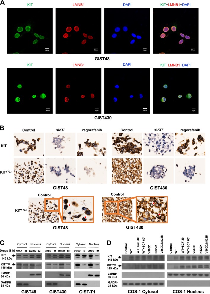
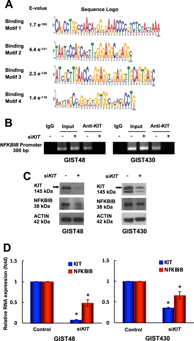
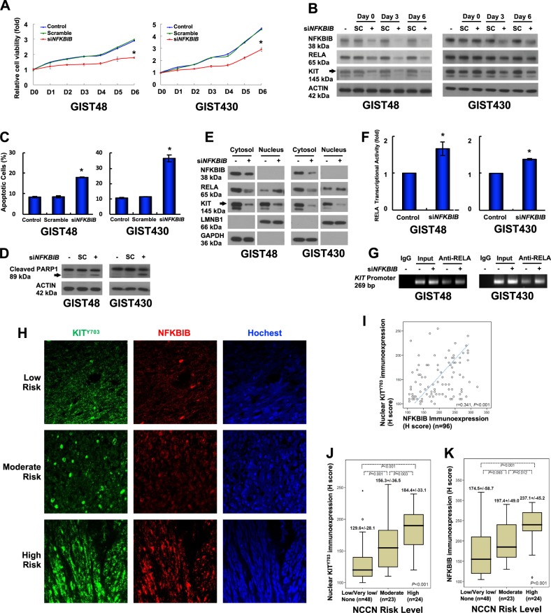
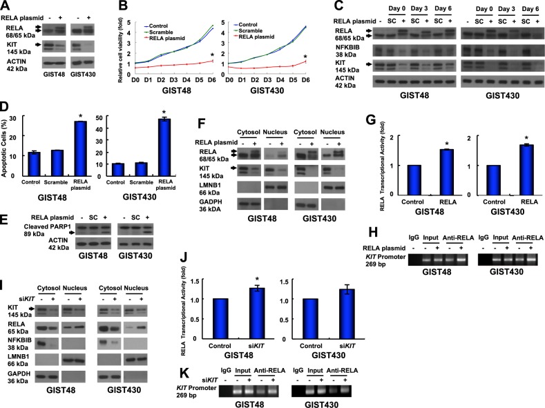
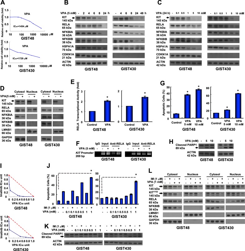
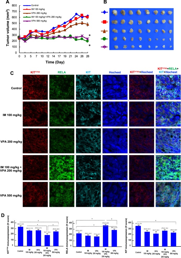
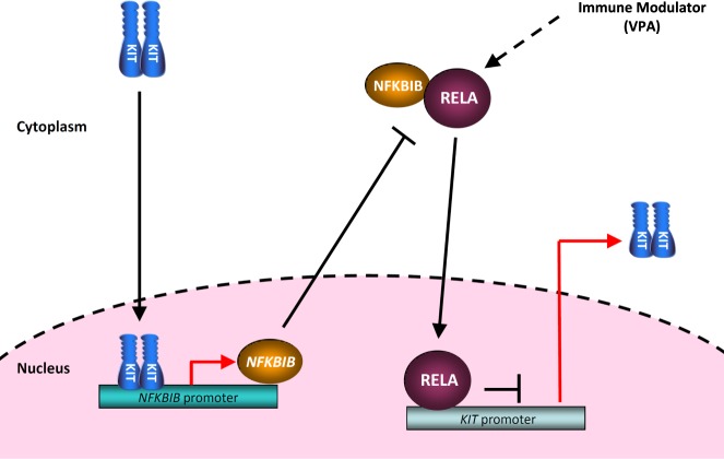
Gastrointestinal stromal tumors (GISTs) are frequently driven by auto-activated, mutant KIT and have durable response to KIT tyrosine kinase inhibitor. However, acquired resistance is an increasing clinical issue in GIST patients receiving front-line imatinib therapy. Our previous studies showed the colocalization of KIT with DAPI-stained nuclei in GIST cells without knowing the role of nuclear KIT in GIST tumorigenesis. In this article, we first identified the binding of nuclear KIT to the promoter of NFKB inhibitor beta (NFKBIB) by chromatin immunoprecipitation (ChIP) sequencing and ChIP assays, which was accompanied with enhanced NFKBIB protein expression in GIST cells. Clinically, high NCCN risk GISTs had significantly higher mean expression levels of nuclear phospho-KIT and NFKBIB as compared with those of intermediate or low/very low-risk GISTs. Conversely, downregulation of NFKBIB by siRNA led to RELA nuclear translocation that could bind to the KIT promoter region and subsequently reduced KIT transcription/expression and the viability of GIST cells. These findings were further confirmed by either RELA overexpression or NFKB/RELA inducer, valproic acid, treatment to result in reduced KIT expression and relative cell viability of imatinib-resistant GIST cells. Combining valproic acid with imatinib showed significantly better growth inhibitory effects on imatinib-resistant GIST48 and GIST430 cells in vitro, and in the GIST430 animal xenograft model. Taken together, these results demonstrate the existence of a nuclear KIT-driven NFKBIB-RELA-KIT autoregulatory loop in GIST tumorigenesis, which are potential targets for developing combination therapy to overcome imatinib-resistant of KIT-expressing GISTs.


Conflict of interest statement

Dr. Li-Tzong Chen reports personal fees from Ono Pharmaceutical, personal fees from Bristol-Myers Squibb, personal fees from Eli Lilly, personal fees from MSD, PharmaEngine, personal fees from Merrimack, grants and personal fees from TTY Biopharm, grants and personal fees from Syncore, personal fees from Five Prime, grants and personal fees from Novartis, personal fees from Pfizer, grants from GlaxoSmithKline, grants from Merck Serono, grants from Polaris, outside the submitted work. The remaining authors declare no potential conflicts of interest.

Figures

Fig. 1
Fig. 1
Distribution of KIT in the cytoplasm and nucleus of GIST cells. a GIST48 and GIST430 cells were stained using antibodies against KIT and LMNB1. After the cells were immunostained, they were visualized by confocal microscopy, and images were acquired through the Cy2, rhodamine, and DAPI channels (×1000). The data were derived from representative images of five fields/picture for each sample. b Cells were transfected with 150 nM siRNA targeting KIT for 72 h or treated with 1 μM regorafenib for 8 h. The cell blocks were analyzed by immunohistochemistry staining against phosphorylated KIT (KITY703) and total KIT. GIST cells were treated with 1 μM IM for 8 h (c), and COS-1 cells were transfected with various KIT mutants and treated with or without SCF for indicated time (d). The cells were fractionated to separate the cytoplasmic and nuclear proteins and analyzed by immunoblotting for KITY703 and total KIT. LMNB1 and GAPDH were used as nuclear and cytoplasmic markers, respectively. All experiments were repeated at least three times
Fig. 2
Fig. 2
Role of nuclear KIT in GIST48 and GIST430 cells. Chromatin obtained from GIST48 cells was cross-linked, sheared, immunoprecipitated using an anti-KIT antibody, and analyzed using next-generation sequencing (NGS). Short reads obtained from NGS were mapped to the reference genome. Enriched reads (compared with a normal IgG-immunoprecipitated control) were adjusted using filters, and their distribution in the genome was determined. a Logos were obtained by running MEME-ChIP with 300-bp summits of the top 600 KIT-bound specific ChIP-seq peaks. The numbers next to the logos indicated the occurrence of the motifs and the statistical significance (E-value). bd GIST48 and GIST430 cells were transfected with 150 nM siRNA targeting KIT for 72 h. b The chromatin was cross-linked, sheared, immunoprecipitated using an anti-KIT antibody, and amplified by PCR. Chromatin that was sheared but not immunoprecipitated was used as an input control. Protein (c) and RNA (d) extracted from parental and KIT-silenced cells were analyzed by immunoblotting and real-time PCR, respectively. Actin served as an internal control for both protein and RNA loading. All experiments were repeated at least three times. The data are expressed as the means ± SD of three or more independent experiments. *p < 0.05
Fig. 3
Fig. 3
Role of KIT-regulated NFKBIB in GIST cell function. a, b GIST48 and GIST430 cells were transfected with 150 nM siRNA targeting NFKBIB or a scrambled control (SC) for 18 h. a The transfected cells were suspended, and equal numbers of cells were seeded into 24-well plates. After 6 h, the cells were attached and examined as the day 0 (D0) control, and the other cells were examined at the indicated times using a relative cell viability assay. b The cells were lysed and analyzed by immunoblotting. Actin served as an internal control. cg Cells were transfected with siRNA targeting NFKBIB or a scrambled control for 72 h. The cells were analyzed by Annexin V staining (c) or immunoblotting against PARP1 (d). e The transfected cells were separated into cytoplasmic and nuclear fractions and analyzed by immunoblotting. LMNB1 and GAPDH were used as nuclear and cytoplasmic markers, respectively. f Nuclear proteins were used to analyze the RELA transcriptional activity. g Chromatin from the transfected cells was cross-linked, sheared, immunoprecipitated using an anti-RELA antibody, and amplified by PCR. Chromatin that was sheared but not immunoprecipitated was used as an input control. All experiments were repeated at least three times. h Representative GIST tissue samples were analyzed by immunostaining against p-KIT (KITY703; green) and NFKBIB (red) and visualized by confocal microscopy. Using photomicrographs, GISTs were classified as low-risk, moderate-risk, or high-risk according to NCCN consensus criteria based on increasing mitoses. i The scatter plot showed the correlations between 96 GIST tissues with various risk levels and the H-scores of the immunoexpression levels of nuclear KITY703 and NFKBIB. The box plot showed the associations between the nuclear KITY703 (j) and NFKBIB (k) immunoexpression levels and the H-scores for tumor grade based on the NCCN risk level according to the Kruskal–Wallis test. The middle line demonstrated the median, the box illustrated the interquartile range, and the whiskers indicated the extreme data points >1.5x the interquartile range from the box. *p < 0.05
Fig. 4
Fig. 4
Roles of RELA in KIT expression and GIST cell function. a Cells were transfected with the RELA/pcDNA3.1 plasmid, lysed, and analyzed by immunoblotting against RELA and KIT. bh The cells were transfected with RELA/pcDNA3.1 or a scrambled control (SC) for 18 h. The transfected cells were resuspended, and equal numbers of cells were seeded into 24-well plates. b After 6 h, the cells were attached and examined as the day 0 (D0) control, and the other cells were examined at the indicated times using a relative cell viability assay. c The cells were lysed and analyzed by immunoblotting. The transfected cells were analyzed by Annexin V staining (d) or immunoblotting against PARP1 (e). f The transfected cells were separated into cytoplasmic and nuclear protein fractions and analyzed by immunoblotting. g The nuclear protein fraction was analyzed to determine RELA transcriptional activity. h Chromatin obtained from the transfected cells was cross-linked, sheared, immunoprecipitated using an anti-RELA antibody, and amplified by PCR. Chromatin that was sheared but not immunoprecipitated was used as an input control. i–k Cells were transfected with siRNA targeting KIT or a scrambled control for 72 h. i The cells were separated into cytoplasmic and nuclear protein fractions and analyzed by immunoblotting. j The nuclear protein fraction was analyzed for RELA transcriptional activity. k Chromatin from the transfected cells was cross-linked, sheared, immunoprecipitated using an anti-RELA antibody, and amplified by PCR. Chromatin that was sheared but not immunoprecipitated was used as an input control. All experiments were repeated at least three times. The data are expressed as the means ± SD of two or more independent experiments. ACTIN (a, c, e) served as an internal control. LMNB1 and GAPDH (f and i) were used as nuclear and cytoplasmic markers, respectively. *p < 0.05
Fig. 5
Fig. 5
Antitumor activity of VPA was mediated by KIT downregulation, and relative cell viability was inhibited by RELA activation. a GIST48 and GIST430 cells were incubated with VPA at the indicated doses, and the IC50 was determined using a relative cell viability assay. bd The cells were treated with 5 mM VPA for the indicated times (b), or treated with the indicated dose of VPA for 24 h (c). The total cell lysates or fractionated proteins (d) were extracted from the cells and analyzed by immunoblotting. e, f The cells were incubated with 5 mM VPA for 24 h. e The nuclear protein fraction was analyzed for RELA transcriptional activity. f Chromatin obtained from VPA-treated cells was cross-linked, sheared, immunoprecipitated using an anti-RELA antibody, and amplified by PCR. Chromatin that was sheared but not immunoprecipitated was used as an input control. g, h The cells were incubated with VPA and then analyzed by Annexin V staining (g) or immunoblotting against PARP1 (h). i Relative cell viability was determined using a methylene blue dye assay as described in the Supplementary Experimental Procedures. The interaction between VPA and IM at the IC50 value was analyzed using the isobologram method. j, k Cells were treated with various concentrations of VPA with or without IM and then analyzed by Annexin V staining (j) or immunoblotting against PARP1 (k). l Cells were treated with 1 mM VPA with or without 1 µM IM for 24 h. The fractionated proteins were extracted from the cells and analyzed by immunoblotting. The data were expressed as the means ± SD of three or more independent experiments. ACTIN (b, c, h, k) served as an internal control. LMNB1 and GAPDH (d and l) were used as nuclear and cytoplasmic markers, respectively. *p < 0.05
Fig. 6
Fig. 6
Effect of VPA either alone or combined with IM in a GIST430 xenograft animal model. ad GIST430 xenografts were established as described in the Materials and Methods section. Mice received an intraperitoneal injection of control (DMSO), 100 mg/kg IM, 200 mg/kg VPA, 100 mg/kg IM with 200 mg/kg VPA, and 500 mg/kg VPA, were administered i.p. twice per week for 4 weeks (n = 8/group). a Tumor volume was calculated as 1/2 x length x width2. b The tumors were collected at the end of drug administration and analyzed by immunostaining (c) against KIT703, RELA, or KIT. The immunoexpression levels were quantified (d). The data are expressed as the means ± SD of three or more independent experiments. *p < 0.001
Fig. 7
Fig. 7
Schematic of the KIT-NFKBIB-RELA autoactivation loop in mutant KIT-expressing GIST cells

Similar articles

Cited by

References

    1. Rubin BP, Heinrich MC, Corless CL. Gastrointestinal stromal tumour. Lancet. 2007;369:1731–41. doi: 10.1016/S0140-6736(07)60780-6. - DOI - PubMed
    1. Nishida T, Hirota S. Biological and clinical review of stromal tumors in the gastrointestinal tract. Histol Histopathol. 2000;15:1293–301. - PubMed
    1. Blanke CD, Demetri GD, von Mehren M, Heinrich MC, Eisenberg B, Fletcher JA, et al. Long-term results from a randomized phase II trial of standard- versus higher-dose imatinib mesylate for patients with unresectable or metastatic gastrointestinal stromal tumors expressing KIT. J Clin Oncol. 2008;26:620–5. doi: 10.1200/JCO.2007.13.4403. - DOI - PubMed
    1. Sleijfer S, Wiemer E, Seynaeve C, Verweij J. Improved insight into resistance mechanisms to imatinib in gastrointestinal stromal tumors: a basis for novel approaches and individualization of treatment. Oncologist. 2007;12:719–26. doi: 10.1634/theoncologist.12-6-719. - DOI - PubMed
    1. Demetri GD, Reichardt P, Kang YK, Blay JY, Rutkowski P, Gelderblom H, et al. Efficacy and safety of regorafenib for advanced gastrointestinal stromal tumours after failure of imatinib and sunitinib (GRID): an international, multicentre, randomised, placebo-controlled, phase 3 trial. Lancet. 2013;381:295–302. doi: 10.1016/S0140-6736(12)61857-1. - DOI - PMC - PubMed

Publication types

MeSH terms

LinkOut - more resources