

Flexor Tendon Injury: Avascular or Vascularized Region Suture? Biomechanical and Histopathological Study in Rabbits
- PMID: 31363280
- PMCID: PMC6597428
- DOI: 10.1055/s-0039-1692458
Flexor Tendon Injury: Avascular or Vascularized Region Suture? Biomechanical and Histopathological Study in Rabbits
Abstract
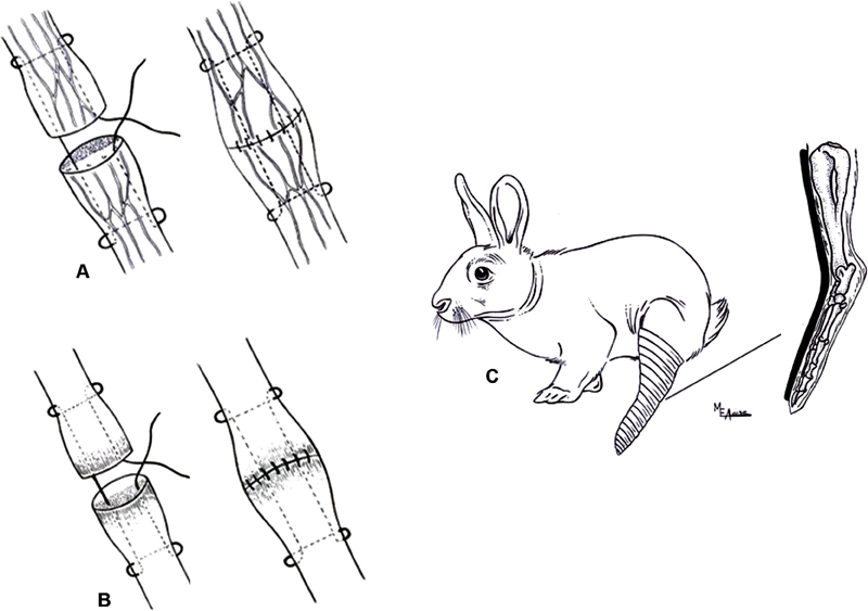
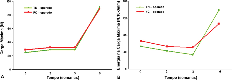
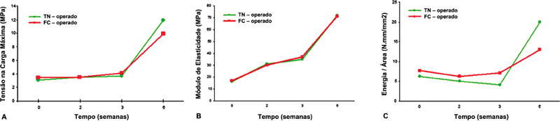
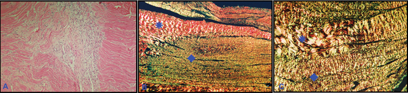
Objectives The present study aims to analyze the mechanical and histopathological aspects of flexor tendon healing focusing on the suture placement site in a vascular or in an avascular region. Methods A total of 83 rabbits were submitted to a Kessler-type central suture in the vascularized tendon region (TN group) and in the avascular tendon region (FC group). The operated limb was immobilized for 3 weeks. The animals were sacrificed in the immediate postoperative period, and at 2, 3 and 6 weeks after the procedure. The mechanical properties studied were: maximum load, stress at maximum load, modulus of elasticity, energy at maximum load, and energy per area. The contralateral tendon was used as control. The histopathological study was descriptive. Results The analysis of the mechanical properties showed similar behavior in both groups, with stabilization or discrete increased values between the immediate period and 3 weeks after the procedure, and marked increased values at 6 weeks. Histopathology demonstrated that the healing process was similar in the TN and FC groups. Conclusion Central suture placement in the vascularized or avascular fibrocartilaginous region results in no differences in the biomechanical and histopathological aspects of flexor tendon healing in rabbits.
Keywords: rabbits; sutures; tendon injuries.
Conflict of interest statement
Figures

Similar articles
-
IMMEDIATE AND LATE EFFECT OF SUTURES IN EXTRASYNOVIAL TENDONS: BIOMECHANICAL STUDY IN RATS.Rev Bras Ortop. 2015 Dec 8;46(3):305-8. doi: 10.1016/S2255-4971(15)30200-7. eCollection 2011 May-Jun. Rev Bras Ortop. 2015. PMID: 27047823 Free PMC article.
-
In Vitro Biomechanical Study on the "Figure-of-Eight" and Kessler Sutures in Swine Flexor Tendons.Rev Bras Ortop (Sao Paulo). 2020 Aug;55(4):445-447. doi: 10.1055/s-0039-1700828. Epub 2020 Jan 9. Rev Bras Ortop (Sao Paulo). 2020. PMID: 32904827 Free PMC article.
-
Effects of PDGF-BB delivery from heparinized collagen sutures on the healing of lacerated chicken flexor tendon in vivo.Acta Biomater. 2017 Nov;63:200-209. doi: 10.1016/j.actbio.2017.09.006. Epub 2017 Sep 7. Acta Biomater. 2017. PMID: 28890257 Free PMC article.
-
Biomechanical evaluation of tendon regeneration with adipose-derived stem cell.J Orthop Res. 2019 Jun;37(6):1281-1286. doi: 10.1002/jor.24182. Epub 2019 Jan 4. J Orthop Res. 2019. PMID: 30474884 Review.
-
[Primary treatment of flexor tendon injuries].Unfallchirurg. 2020 Feb;123(2):89-96. doi: 10.1007/s00113-019-00762-w. Unfallchirurg. 2020. PMID: 31970427 Review. German.
References
-
- Kleinert H E, Smith D J., Jr . Baltimore: Williams & Wilkins; 1991. Primary and secondary repairs of flexor and extensor tendon injuries; pp. 241–261.
-
- Culp R W, Taras J S. St. Louis: Mosby; 1995. Primary care of flexor tendon injuries; pp. 417–431.
-
- Aoki M, Manske P R, Pruitt D L, Kubota H, Larson B J. Work of flexion after flexor tendon repair according to the placement of sutures. Clin Orthop Relat Res. 1995;(320):205–210. - PubMed
-
- Soejima O, Diao E, Lotz J C, Hariharan J S. Comparative mechanical analysis of dorsal versus palmar placement of core suture for flexor tendon repairs. J Hand Surg Am. 1995;20(05):801–807. - PubMed
-
- Komanduri M, Phillips C S, Mass D P. Tensile strength of flexor tendon repairs in a dynamic cadaver model. J Hand Surg Am. 1996;21(04):605–611. - PubMed
LinkOut - more resources
Full Text Sources