. 2019 Mar;58(1):13-22.
doi: 10.20471/acc.2019.58.01.02.

SONOELASTOGRAPHIC FEATURES OF HIGH-RISK BREAST LESIONS AND DUCTAL CARCINOMA IN SITU - A PILOT STUDY

Affiliations

SONOELASTOGRAPHIC FEATURES OF HIGH-RISK BREAST LESIONS AND DUCTAL CARCINOMA IN SITU - A PILOT STUDY

Maja Crnogorac et al. Acta Clin Croat. 2019 Mar.

Abstract

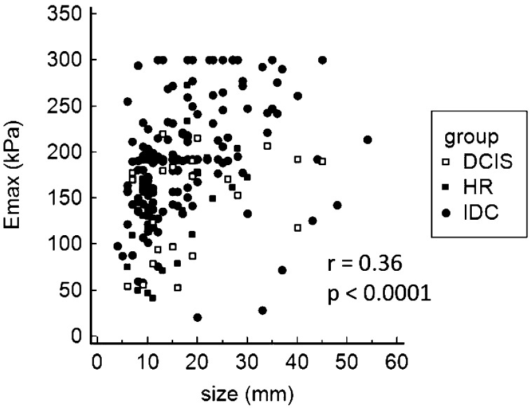
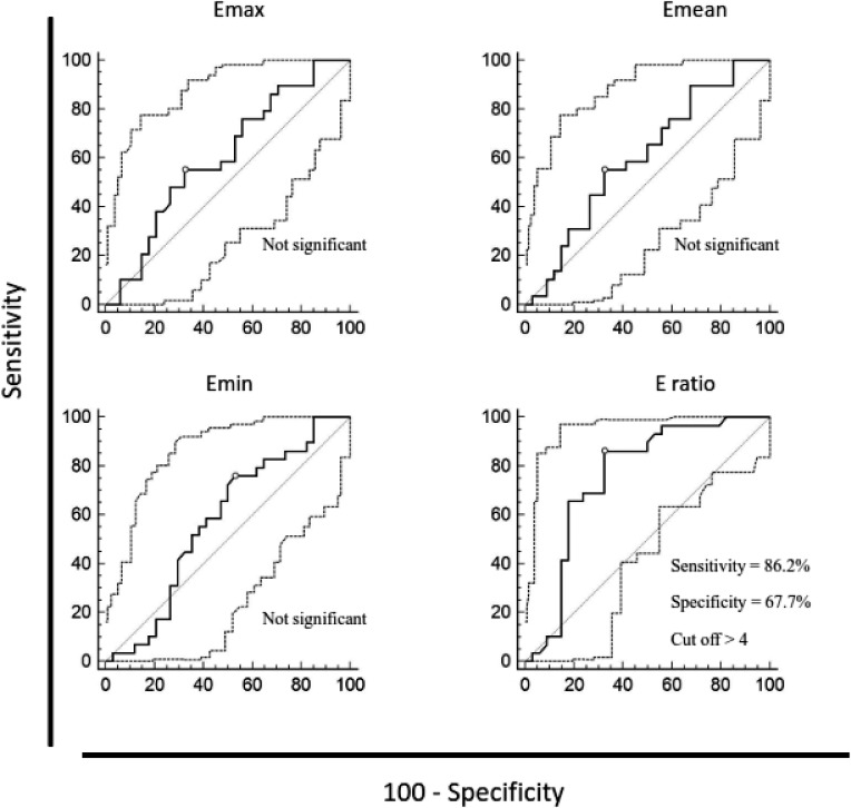
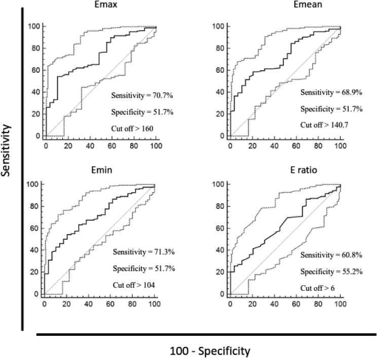
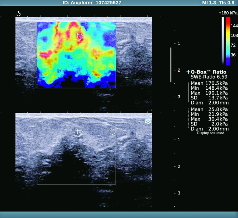
The aim of this study was to evaluate the quantitative sonoelastographic values recorded on shear-wave sonoelastography (SWE) of high-risk breast lesions and ductal carcinoma in situ (DCIS). We retrospectively analyzed histopathologic and SWE data (quantitative maximum, minimum and mean stiffness, lesion-to-fat ratio (E-ratio), lesion size) of 228 women referred to our Department for core needle breast biopsy during a four-year period. Among 230 lesions, histopathologic findings showed 34 high-risk breast lesions and 29 DCIS, which were compared with 167 ductal invasive carcinomas. High-risk lesions had lower values of all sonoelastographic features than ductal in situ and invasive carcinoma, however, only E-ratio showed a statistically significant difference in comparison to DCIS (3.7 vs. 6, p<0.001). All sonoelastographic features showed significant difference between in situ and invasive carcinoma. There was a significant correlation between lesion size and stiffness (r=0.36; p<0.001). Stiffness measured by SWE is an effective predictor of the histopathologic severity of sonographically detectable breast lesions. Elasticity values of high-risk lesions are significantly lower than those of malignant lesions. Furthermore, we showed that along with the sonographic appearance, which in most cases shows typical microcalcifications, DCIS had significantly different elasticity parameters than invasive carcinoma.

Keywords: Breast neoplasms – classification; Breast neoplasms – diagnostic imaging; Carcinoma, intraductal; Elastic modulus; Elasticity imaging techniques; Sensitivity and specificity; Shear-wave elastography (SWE); Ultrasonography, mammary.


Figures

Fig. 1
Fig. 1
Correlation of maximum lesion diameter (mm) and maximum lesion stiffness (kPa). HRL = high risk lesions; DCIS = ductal carcinoma in situ; IDC = invasive ductal carcinoma; Emax = maximum stiffness
Fig. 2
Fig. 2
Discriminatory ability of maximum, mean and minimum lesion stiffness and lesion-to-fat stiffness ratio between high-risk breast lesions and ductal carcinoma in situ, according to receiver operating characteristic (ROC) curves. Diagnostic efficacy for those values was assessed using the sensitivity and specificity at the specified cut-off point. ROC curve analysis, p values = 0.05 were considered statistically significant. Emax = maximum stiffness; Emean = mean stiffness; Emin = minimum stiffness; E-ratio = lesion-to-fat ratio
Fig. 3
Fig. 3
Discriminatory ability of maximum, mean and minimum lesion stiffness and lesion-to-fat stiffness ratio between ductal carcinoma in situ and ductal invasive carcinoma, according to receiver operating characteristic (ROC) curves. Diagnostic efficacy for those values was assessed using the sensitivity and specificity at the specified cut-off point. ROC curve analysis, p values = 0.05 were considered statistically significant. Emax = maximum stiffness; Emean = mean stiffness; Emin = minimum stiffness; E-ratio = lesion-to-fat ratio
Fig. 4
Fig. 4
Ductal in situ carcinoma – sonoelastographic and gray-scale ultrasound image.

Similar articles

Cited by

References

    1. Croatian Institute for Public Health, Croatian National Cancer Registry. Bulletin no.38: Cancer incidence in Croatia 2013.
    1. Berg WA, Gutierrez L, NessAiver MS, Carter WB, Bhargavan M, Lewis RS, et al. Diagnostic accuracy of mammography, clinical examination, US, and MR imaging in preoperative assessment of breast cancer. Radiology. 2004;233:830–49. 10.1148/radiol.2333031484 - DOI - PubMed
    1. Stavros AT, Thickman D, Rapp CL, Dennis MA, Parker SH, Sisney GA. Solid breast nodules: use of sonography to distinguish between benign and malignant lesions. Radiology. 1995;196:123–34. 10.1148/radiology.196.1.7784555 - DOI - PubMed
    1. Çebi Olgun D, Korkmazer B, Kilic F, Dikici AS, Velidedeoglu M, Aydoga F, et al. Use of shear wave elastography to differentiate benign and malignant breast lesions. Diagn Interv Radiol. 2014;20:239–44. 10.5152/dir.2014.13306 - DOI - PMC - PubMed
    1. Athanasiou A, Tardivon A, Tanter M, Sigal-Zafrani B, Bercoff J, Deffieux T, et al. Breast lesions: quantitative elastography with supersonic shear imaging – preliminary results. Radiology. 2010;256:297–303. 10.1148/radiol.10090385 - DOI - PubMed

MeSH terms

LinkOut - more resources