. 2018 Dec;16(4):116-130.
doi: 10.1007/s12018-018-9253-0. Epub 2018 Dec 6.

Systemic Bone Loss After Fracture

Affiliations

Systemic Bone Loss After Fracture

Benjamin Osipov et al. Clin Rev Bone Miner Metab. 2018 Dec.

Abstract

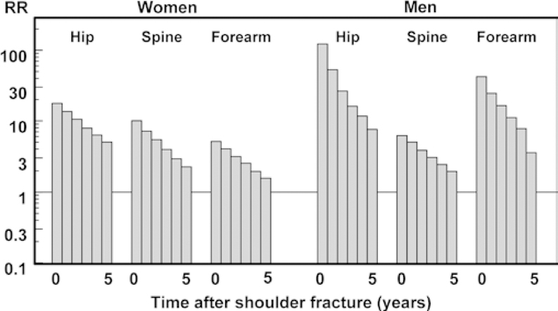
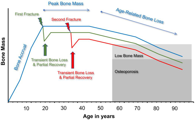
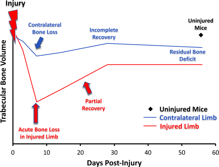
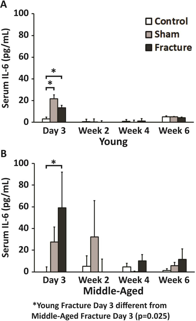
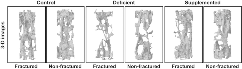
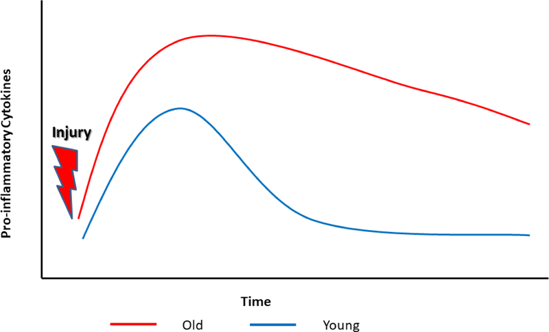
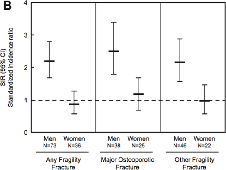
A history of prior fracture is the most reliable indicator of prospective fracture risk. Increased fracture risk is not confined to the region of the prior fracture, but is operant at all skeletal sites, providing strong evidence of systemic bone loss after fracture. Animal and human studies suggest that systemic bone loss begins shortly after fracture and persists for several years in humans. In fact, bone quantity and bone quality may never fully return to their pre-fracture levels, especially in older subjects, demonstrating a need for improved understanding of the mechanisms leading to systemic bone loss after fracture in order to reduce subsequent fracture risk. Although the process remains incompletely understood, mechanical unloading (disuse), systemic inflammation, and hormones that control calcium homeostasis may all contribute to systemic bone loss. Additionally, individual factors can potentially affect the magnitude and time course of systemic bone loss and recovery. The magnitude of systemic bone loss correlates positively with injury severity and age. Men may also experience greater bone loss or less recovery than women after fracture. This review details the current understanding of systemic bone loss following fracture, including possible underlying mechanisms and individual factors that may affect this injury response.

Keywords: Bone Loss; Fracture Risk; Fracture healing; Osteoporosis.


Figures

Fig. 1
Fig. 1
Relative Risk (RR) of hip, spine, and forearm fracture for 5 years after a shoulder fracture in men and women aged 60 years [3]
Fig. 2
Fig. 2
Proposed lifetime trajectory of bone mass. Fractures may cause a transient loss of bone that is not fully recovered, resulting in an altered bone mass trajectory and an increased risk of future fractures
Fig. 3
Fig. 3
Trabecular bone loss post-injury. Injury causes acute bone loss followed by an incomplete recovery in both the injured and contralateral limb
Fig. 4
Fig. 4
Enzyme-linked immunosorbent assay (ELISA) of serum interleukin-6 levels in (A) young and (B) middle-aged mice after fracture. At day 3, young Fractured mice had a 3.5 fold increase in serum IL-6, while middle-aged Fractured mice had a 21.9 fold (p<0.001) increase in serum IL-6 compared to age-matched Control mice. Error bars represent standard deviation. * denotes p≤0.05 [34]
Fig. 5
Fig. 5
μCT images of lumbar vertebrae from non-fractured and fractured ovariectomized mice fed a control, Calcium/ Vitamin D-deficient, or Calcium/ Vitamin D- supplemented diet. Fractured Calcium/ Vitamin D-deficient mice exhibited the greatest bone loss post-fracture compared to non-fractured controls from the same dietary group [33]
Fig. 6
Fig. 6
Changes in pro-inflammatory cytokine concentrations post-fracture in young and old individuals. Prior to fracture, old people have higher baseline pro-inflammatory cytokine level. After fracture cytokine levels reach a higher peak in old people and take longer to decrease [121]
Fig. 7
Fig. 7
Standardized incidence ratio (SIR) for the risk of fracture at age ≥50 years for men and women following a distal forearm fracture in childhood (age ≤18 years) from 1935–1992 [10]

Similar articles

Cited by

References

    1. Learn What Osteoporosis Is and What It’s Caused by [Internet]. Natl. Osteoporos. Found. [cited 2018 Jul 27]. Available from: https://www.nof.org/patients/what-is-osteoporosis/
    1. Christiansen BA, Harrison SL, Fink HA, Lane NE, Study of Osteoporotic Fractures Research Group. Incident fracture is associated with a period of accelerated loss of hip BMD: the study of osteoporotic fractures. Osteoporos Int. 2018;1–9. - PMC - PubMed
    1. Johnell O, Kanis JA, Odén A, Sernbo I, Redlund-Johnell I, Petterson C, et al. Fracture risk following an osteoporotic fracture. Osteoporos Int. 2004;15:175–9. - PubMed
    1. Ahmed LA, Center JR, Bjørnerem Å, Bluic D, Joakimsen RM, Jørgensen L, et al. Progressively increasing fracture risk with advancing age after initial incident fragility fracture: the Tromsø study. J Bone Miner Res. 2013;28:2214–21. - PubMed
    1. Clinton J, Franta A, Polissar NL, Neradilek B, Mounce D, Fink HA, et al. Proximal humeral fracture as a risk factor for subsequent hip fractures. J Bone Joint Surg Am. 2009;91:503–11. - PMC - PubMed