

Improving diabetic and hypertensive retinopathy with a medical food containing L-methylfolate: a preliminary report
- PMID: 31363484
- PMCID: PMC6643316
- DOI: 10.1186/s40662-019-0147-0
Improving diabetic and hypertensive retinopathy with a medical food containing L-methylfolate: a preliminary report
Abstract
Background: Homocysteine and vitamin D may play a role in the development of diabetic and hypertensive retinopathy in patients with diabetes mellitus (DM) and hypertension. Supplementing food with L-methylfolate and vitamin D theoretically may improve diabetic and hypertensive retinopathy, however, the outcome of these nutritional approaches has not been fully examined. A retrospective case review was done of cases of retinopathy reversal in patients on Ocufolin™ and a similar nonprescription multivitamin, Eyefolate™. In this study, they were administered L-methylfolate (2.7 mg and 3.0 mg, respectively) and vitamin D3 (4500 IU each). These dosages are significantly above the RDA but well below levels associated with toxicity.
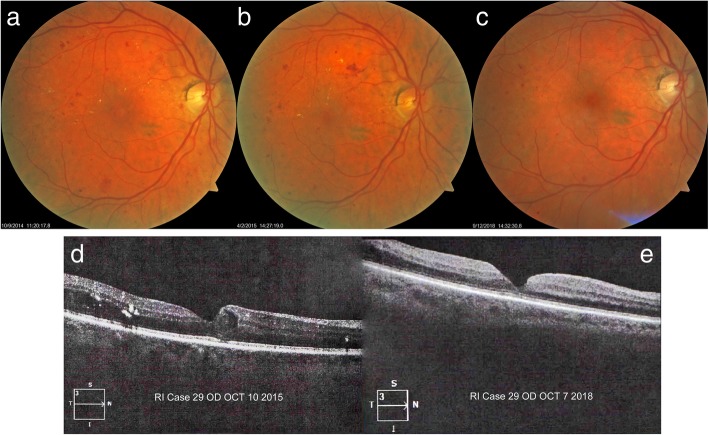
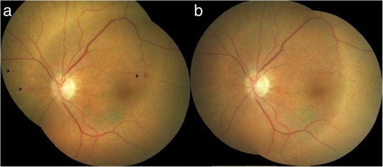
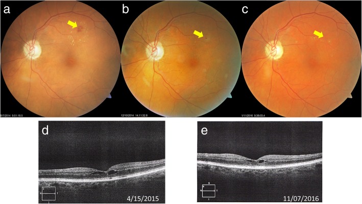
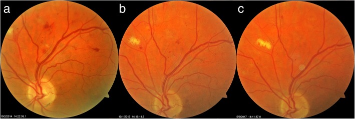
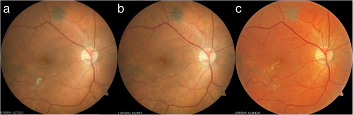
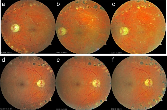
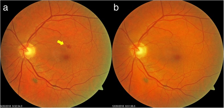
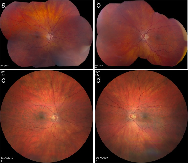
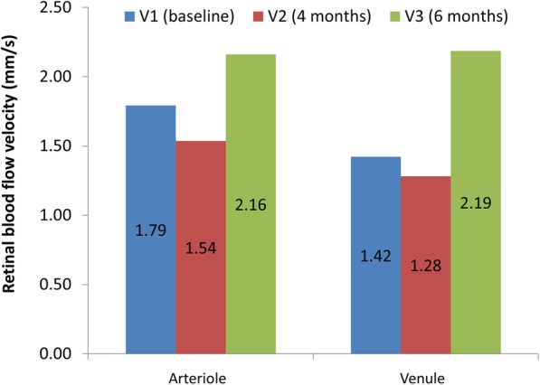
Case presentation: Seven patients had nonproliferative diabetic retinopathy (NPDR) and some of them had hypertension. One patient had only hypertensive retinopathy. All patients were instructed to take Ocufolin™ medical food as a food supplement. Baseline genetic testing for MTHFR polymorphisms was conducted. Fundus photography was used to document the fundus condition of the enrolled eyes in 8 NPDR patients at the initial and follow-up visits. Microaneurysms (MA) and exudates were observed to be improved in some trial patients. All subjects had one or more MTHFR polymorphisms. All had diabetic retinopathy, hypertensive retinopathy, or both. MAs were resolved, and exudates were decreased in 8/8 cases after taking the medical food. Retinal edema was found in 2/8 cases and improved or resolved in both cases after taking the medical food or the supplement. The best corrected visual activity was stable or improved in 8/8 cases.
Conclusion: We report a series of diabetic and hypertensive retinopathy cases with MTHFR polymorphisms and the improvement of retinal microvasculature (mainly MAs) in serial fundus photography after taking a medical food or supplement containing L-methylfolate and vitamin D. It appears that the use of nutritional supplements and medical foods containing L-methylfolate and vitamin D may be effective in facilitating the improvement of diabetic and hypertensive retinopathy.
Keywords: Diabetes; Homocysteine; L-methylfolate; MTHFR C677T; Microaneurysms; Retinopathy; Vitamin D.
Conflict of interest statement
Competing interestsDr. Brown holds an ownership interest in Global Healthcare Focus, a small research and development company concerned with developing nutritional products to improve health. Other authors have no competing interests.
Figures
Similar articles
-
Nutritional and medical food therapies for diabetic retinopathy.Eye Vis (Lond). 2020 Jun 18;7:33. doi: 10.1186/s40662-020-00199-y. eCollection 2020. Eye Vis (Lond). 2020. PMID: 32582807 Free PMC article. Review.
-
Improved Retinal Microcirculation in Mild Diabetic Retinopathy Patients Carrying MTHFR Polymorphisms Who Received the Medical Food, Ocufolin®.Clin Ophthalmol. 2022 May 16;16:1497-1504. doi: 10.2147/OPTH.S358753. eCollection 2022. Clin Ophthalmol. 2022. PMID: 35607436 Free PMC article.
-
Effects of Methylenetetrahydrofolate Reductase (MTHFR) Polymorphisms on Retinal Tissue Perfusion in Mild Diabetic Retinopathy Patients Receiving the Medical Food, Ocufolin®.Clin Ophthalmol. 2023 Apr 12;17:1121-1127. doi: 10.2147/OPTH.S401743. eCollection 2023. Clin Ophthalmol. 2023. PMID: 37077224 Free PMC article.
-
Improved conjunctival microcirculation in diabetic retinopathy patients with MTHFR polymorphisms after Ocufolin™ Administration.Microvasc Res. 2020 Nov;132:104066. doi: 10.1016/j.mvr.2020.104066. Epub 2020 Aug 27. Microvasc Res. 2020. PMID: 32860770 Free PMC article.
-
Three-year follow-up study of blood-retinal barrier and retinal thickness alterations in patients with type 2 diabetes mellitus and mild nonproliferative diabetic retinopathy.Arch Ophthalmol. 2004 Feb;122(2):211-7. doi: 10.1001/archopht.122.2.211. Arch Ophthalmol. 2004. PMID: 14769598
Cited by
-
Nutritional and medical food therapies for diabetic retinopathy.Eye Vis (Lond). 2020 Jun 18;7:33. doi: 10.1186/s40662-020-00199-y. eCollection 2020. Eye Vis (Lond). 2020. PMID: 32582807 Free PMC article. Review.
-
The emerging role of medical foods and therapeutic potential of medical food-derived exosomes.Nanoscale Adv. 2023 Nov 11;6(1):32-50. doi: 10.1039/d3na00649b. eCollection 2023 Dec 19. Nanoscale Adv. 2023. PMID: 38125597 Free PMC article. Review.
-
Improved Retinal Microcirculation in Mild Diabetic Retinopathy Patients Carrying MTHFR Polymorphisms Who Received the Medical Food, Ocufolin®.Clin Ophthalmol. 2022 May 16;16:1497-1504. doi: 10.2147/OPTH.S358753. eCollection 2022. Clin Ophthalmol. 2022. PMID: 35607436 Free PMC article.
-
Case series of retinal vein occlusions showing early recovery using oral l-methylfolate.Ther Adv Ophthalmol. 2024 Apr 15;16:25158414241240687. doi: 10.1177/25158414241240687. eCollection 2024 Jan-Dec. Ther Adv Ophthalmol. 2024. PMID: 38628356 Free PMC article.
-
Prevalence and risk factors of diabetic retinopathy among Chinese adults with type 2 diabetes in a suburb of Shanghai, China.PLoS One. 2022 Oct 4;17(10):e0275617. doi: 10.1371/journal.pone.0275617. eCollection 2022. PLoS One. 2022. PMID: 36194621 Free PMC article.
References
-
- https://nei.nih.gov/eyedata/diabetic. Accessed 18 June 2019.
-
- http://www.cdc.gov/media/releases/2017/p0718-diabetes-report.html. Accessed 18 June 2019.
Publication types
LinkOut - more resources
Full Text Sources
Other Literature Sources
