. 2019 Nov 15;28(22):3742-3754.
doi: 10.1093/hmg/ddz188.

AAV9-Stathmin1 gene delivery improves disease phenotype in an intermediate mouse model of spinal muscular atrophy

Affiliations

AAV9-Stathmin1 gene delivery improves disease phenotype in an intermediate mouse model of spinal muscular atrophy

E Villalón et al. Hum Mol Genet. .

Abstract

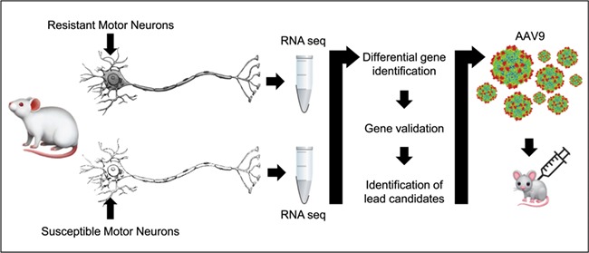
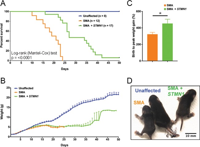
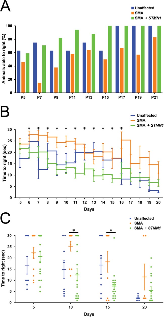
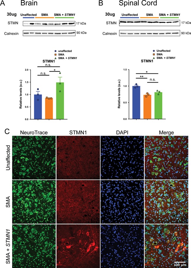
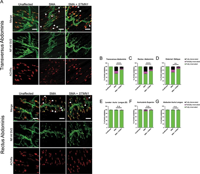
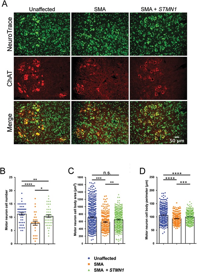
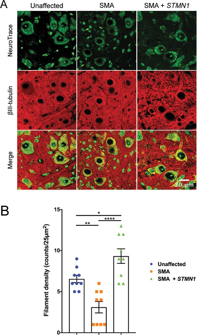
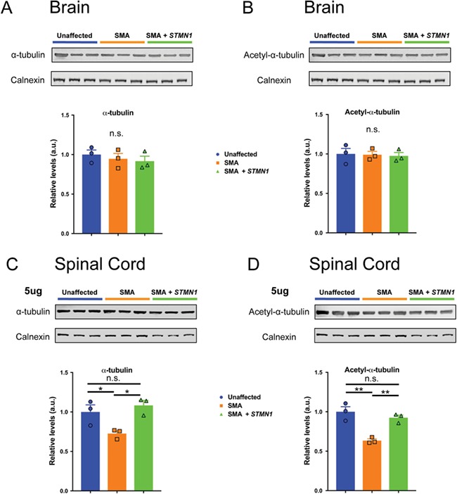
Spinal muscular atrophy (SMA) is a devastating infantile genetic disorder caused by the loss of survival motor neuron (SMN) protein that leads to premature death due to loss of motor neurons and muscle atrophy. The approval of an antisense oligonucleotide therapy for SMA was an important milestone in SMA research; however, effective next-generation therapeutics will likely require combinatorial SMN-dependent therapeutics and SMN-independent disease modifiers. A recent cross-disease transcriptomic analysis identified Stathmin-1 (STMN1), a tubulin-depolymerizing protein, as a potential disease modifier across different motor neuron diseases, including SMA. Here, we investigated whether viral-based delivery of STMN1 decreased disease severity in a well-characterized SMA mouse model. Intracerebroventricular delivery of scAAV9-STMN1 in SMA mice at P2 significantly increased survival and weight gain compared to untreated SMA mice without elevating Smn levels. scAAV9-STMN1 improved important hallmarks of disease, including motor function, NMJ pathology and motor neuron cell preservation. Furthermore, scAAV9-STMN1 treatment restored microtubule networks and tubulin expression without affecting tubulin stability. Our results show that scAAV9-STMN1 treatment improves SMA pathology possibly by increasing microtubule turnover leading to restored levels of stable microtubules. Overall, these data demonstrate that STMN1 can significantly reduce the SMA phenotype independent of restoring SMN protein and highlight the importance of developing SMN-independent therapeutics for the treatment of SMA.


Figures

Figure 1
Figure 1
Strategy for identifying potential disease gene modifiers. The first step is to identify pools of motor neurons that are resistant and vulnerable to pathogenesis, which is determined by the vulnerability to denervation of target muscle fibers. Then by laser microdissection, motor neuron cell bodies are isolated and RNA is extracted from both pools. Transcriptomic analysis is performed on both pools, and differentially expressed genes are identified and validated to generate a list of potential genes. These genes are then packaged into AAV9 virus and used to treat neonatal mouse models of the disease via ICV or IV injection. The effects of the gene modifier are then evaluated.
Figure 2
Figure 2
scAAV9-STMN1 treatment increases lifespan and birth-to-peak weight gain in SMA mice. Phenotypic analyses in SMA mice after ICV injection of 1.0 × 1011 scAAV9-STMN1 viral injection at P2. (A) Survival was significantly increased (P < 0.001) in treated SMA mice compared to untreated SMA, healthy controls survived past 50 days. (B) Average weight gain was not different in treated SMA mice compared to untreated SMA up to day 25. (C) Birth-to-peak weight gain was significantly increased (P = 0.0486) in treated SMA mice compared to untreated SMA. (D) Representative image showing health pup at P25 and improvement in overt appearance of treated SMN mice compared to untreated SMA mice. N = 8, healthy control; n = 13, untreated SMA; n = 17, treated SMA. Data expressed as mean ± SEM.
Figure 3
Figure 3
Motor performance was improved in SMA mice following STMN1 treatment. TTR analysis in unaffected, untreated SMA and scAAV9-STMN1-treated SMA mice from P5 to P25. (A) Percent of animals able to right at each specified time point shows an improvement of treated SMA mice compared to untreated SMA mice. (B) Daily average TTR (s) shows that treated SMA mice are able to right significantly faster than untreated SMA mice from Days P6 to P16 (P < 0.05). Moreover, SMA mice treated from P17 to P20 perform better than untreated SMA mice, although not statistically significantly. Moreover, no statistical significance was found between unaffected and treated SMA mice from P17 to P20. TTR comparisons were analyzed by a two-way-ANOVA followed by a Holm–Sidak post hoc test for multiple comparisons. *P < 0.05. N = 8, healthy control; n = 13, untreated SMA; n = 17, treated SMA. Data expressed as mean ± SEM.
Figure 4
Figure 4
scAAV9-STMN1 treatment results in a significant increase in STMN1 protein expression in peripheral tissues of SMA mice. Western blot analysis from brain tissue show that ICV injection of scAAV9-STMN1 leads to significant upregulation of STMN1 protein expression. (A) Western blots show upregulation of STMN1 protein in treated SMA mice compared to untreated SMA mice. (B) Quantification of blots demonstrate a significant increase in STMN1 protein expression in treated SMA mice compared to untreated mice in peripheral tissues. Comparisons were analyzed by Student t-test. *P < 0.05. Data expressed as mean ± SEM. n = 3 animals per treatment.
Figure 5
Figure 5
scAAV9-STMN1 treatment improves NMJ pathology in vulnerable muscles of SMA mice. Immunohistochemistry analysis of NMJ innervation in vulnerable (TVA, RA and EO) and resistant (levator auris longus (rostral), AS and AAL) muscles from unaffected, untreated SMA and STMN1-treated SMA mice. Muscles were immunostained to label the axon (NF-H), axon terminal (SV) and endplate (AChRs). (A) Representative images of PND18 TVA and RA (vulnerable) muscles showing reduced frequency of denervated endplates in scAAV9-STMN1-treated SMA mice (right panel) compared to untreated SMA mice (middle panel). Unaffected controls show no denervated endplates (left panel). Maximum projection confocal microscope images taken at ×20 magnification. White arrows point to the location of denervated endplates. (B, C and D) Quantification of NMJ denervation showing percentages of fully innervated, partially innervated and fully denervated endplates in vulnerable muscles. (E, F and G) Quantification of NMJ denervation in resistant muscles showing percent fully innervated, partially innervated and fully denervated NMJs. Denervation analysis shows that STMN1 treatment increases frequency of fully innervated endplates in SMA vulnerable muscles without affecting innervation of resistant muscles. For ease of presentation, only statistical comparisons of fully innervated NMJ percentages are presented. Data analyzed by a two-way ANOVA followed by a Tukey post hoc test for multiple comparisons. Data expressed as mean ± SEM. ****P < 0.0001, ***P < 0.001, n.s. = not significant. n = 3 animals per treatment.
Figure 6
Figure 6
scAAV9-STMN1 treatment prevents motor neuron cell body pathology in SMA mice. Cytological analysis of motor neuron cell body in L3–L5 spinal cord from PND18 treatment groups. (A) Representative images of unaffected (left panel), untreated SMA (middle panel) and treated SMA (right panel) lumbar motor neurons immunostained with Nissl stain (neurotrace) to label neuronal cell bodies and ChAT to label specifically motor neurons. ChAT stain revealed an increased number of motor neurons in STMN1-treated SMA compared to untreated SMA mice. Fluorescent microscope images taken at ×40 magnification. (B) Quantification of motor neuron cell bodies showed a significant increase in cell numbers in STMN1-treated SMA mice compared to untreated SMA. However, STMN1-treated motor neuron cell numbers were still significantly lower than unaffected healthy controls. (C) Morphometric analysis showed a significant improvement in motor neuron cell body area (μm2) of STMN1-treated SMA mice compared to untreated SMA mice. (D) Analysis of cell body perimeter also showed significant improvement in SMTN1-treated SMA motor neurons as compared to untreated SMA. However, these improvements were still significantly lower than unaffected control. Data were analyzed by a one-way ANOVA followed by a Tukey post hoc test for multiple comparisons. Data expressed as mean ± SEM. ****P < 0.0001, ***P < 0.001, **P < 0.01, *P < 0.05, n.s. = not significant. n = 3 animals per treatment (n > 400, cells measured per treatment).
Figure 7
Figure 7
scAAV9-STMN1 treatment restores microtubule filamentous networks in SMA mice. Tubulin filament immunohistochemistry from L3–L5 spinal cords of treatment groups. (A) Representative images of lumbar spinal cord ventral horn motor neurons stained with Nissl stain (neurotrace) and anti-BIII-tubulin antibodies conjugated to Alexa Fluor 594 to label microtubule filaments. Unaffected control tissues showed distinct filamentous networks (left panel), which were absent in untreated SMA ventral horn spinal cords (middle panel). STMN1 treatment restored filamentous networks in SMA lumbar spinal cords to a greater extent compared to unaffected controls. Maximum projection high-resolution confocal microscope images taken at ×63 magnification. (B) Quantifications of filaments per 25 μm2 showed a significant increase in filament density in treated SMA spinal cord compared to untreated and unaffected controls. Data were analyzed by a one-way ANOVA followed by a Tukey post hoc test for multiple comparisons. Data expressed as mean ± SEM. ****P < 0.0001, **P < 0.01, *P < 0.05, n.s. = not significant.
Figure 8
Figure 8
scAAV9-STMN1 treatment restores microtubule levels without affecting microtubule stability in the spinal cord of SMA mice. Western blot analyses of tubulin expression and tubulin stability in brain and spinal cord of PND8-unaffected, untreated SMA and STMN1-treated SMA mice. (A and B) Protein extracts from brain immunoblotted with antibodies specific for α-tubulin (A, top) and acetylated-α-tubulin (B, top) and blot quantifications showing no difference in α-tubulin (A, bottom) or acetylated-α-tubulin (B, bottom) expression in untreated SMA or treated SMA mice compared to unaffected controls. (C and D) Protein extracts form lumbar spinal cord immunoblotted for α-tubulin (C, bottom) and acetylated-α-tubulin (D, top) and quantifications showed significant decrease in α-tubulin (C, bottom) and acetylated-α-tubulin (D, bottom) in untreated SMA spinal cords compared to unaffected controls. Moreover, quantifications show restoration of both α-tubulin and acetylated-α-tubulin (C and D, bottom) in STMN1-treated SMA spinal cords compared to unaffected controls. Data were analyzed by a one-way ANOVA followed by a Tukey post hoc test for multiple comparisons. Data expressed as mean ± SEM. **P < 0.01, *P < 0.05, n.s. = not significant. n = 3 animals per treatment.

Similar articles

Cited by

References

    1. Lefebvre S., Burglen L., Reboullet S., Clermont O., Burlet P., Viollet L., Benichou B., Cruaud C., Millasseau P., Zeviani M. et al. (1995) Identification and characterization of a spinal muscular atrophy-determining gene. Cell, 80, 155–165. - PubMed
    1. Kolb S.J. and Kissel J.T. (2011) Spinal muscular atrophy: a timely review. Arch. Neurol., 68, 979–984. - PMC - PubMed
    1. Burghes A.H. and Beattie C.E. (2009) Spinal muscular atrophy: why do low levels of survival motor neuron protein make motor neurons sick? Nat. Rev. Neurosci., 10, 597–609. - PMC - PubMed
    1. Lorson C.L., Hahnen E., Androphy E.J. and Wirth B. (1999) A single nucleotide in the SMN gene regulates splicing and is responsible for spinal muscular atrophy. Proc. Natl. Acad. Sci. U.S.A., 96, 6307–6311. - PMC - PubMed
    1. Monani U.R., Lorson C.L., Parsons D.W., Prior T.W., Androphy E.J., Burghes A.H. and McPherson J.D. (1999) A single nucleotide difference that alters splicing patterns distinguishes the SMA gene SMN1 from the copy gene SMN2. Hum. Mol. Genet., 8, 1177–1183. - PubMed

Publication types

MeSH terms