Review
. 2019 Nov;68(11):915-932.
doi: 10.1007/s00011-019-01273-5. Epub 2019 Jul 30.

The effect of palmitic acid on inflammatory response in macrophages: an overview of molecular mechanisms

Affiliations
Review

The effect of palmitic acid on inflammatory response in macrophages: an overview of molecular mechanisms

Jan Korbecki et al. Inflamm Res. 2019 Nov.

Abstract

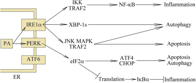
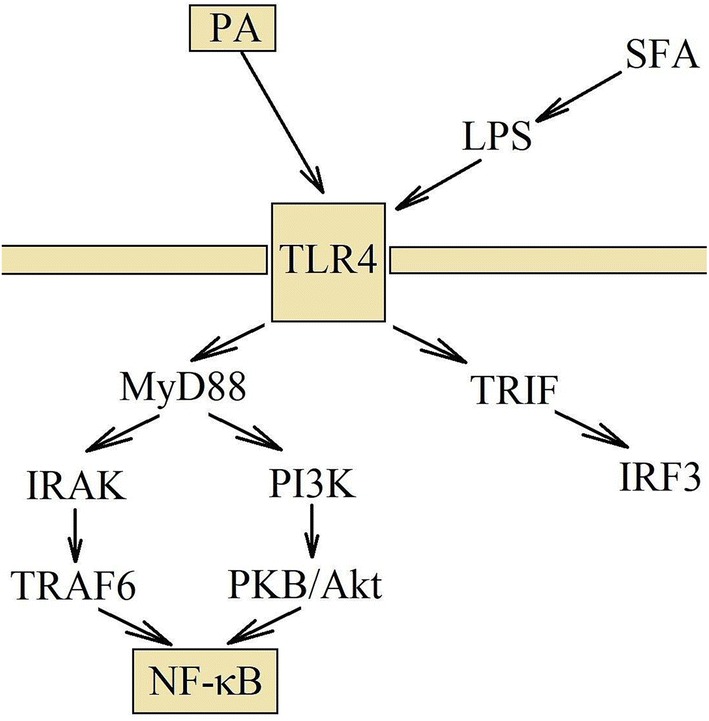
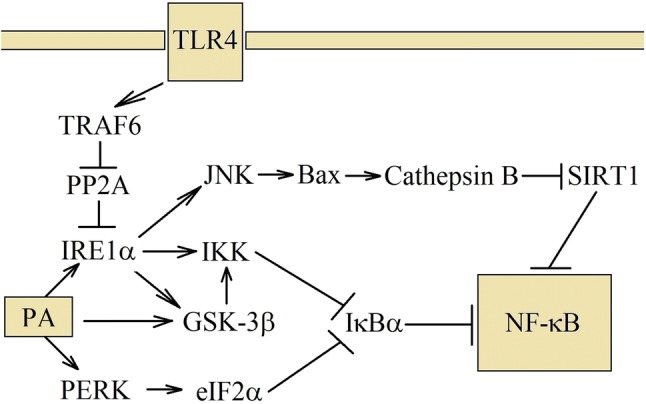
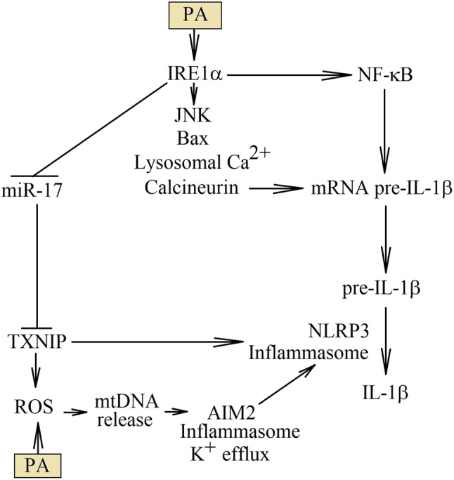
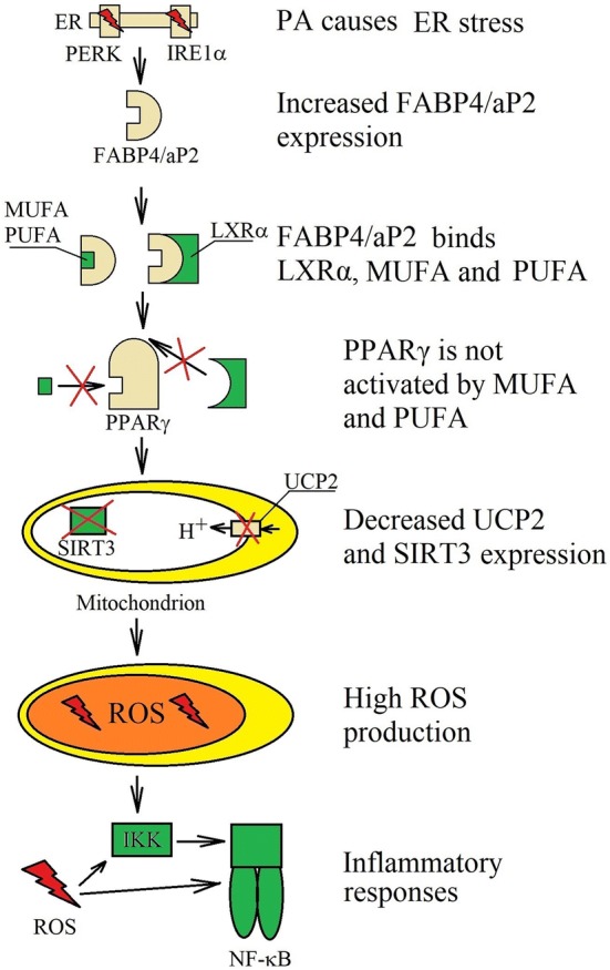
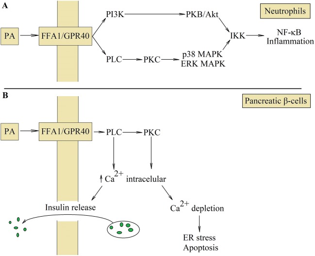
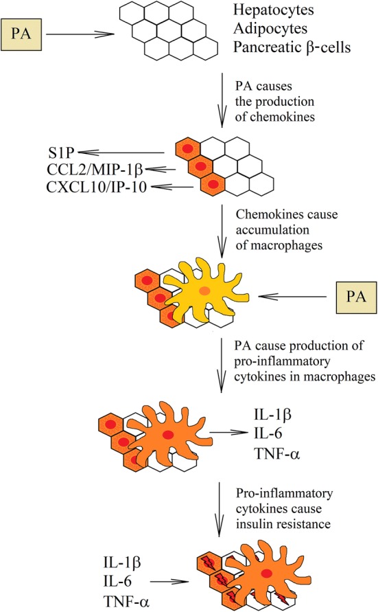
Palmitic acid is a saturated fatty acid whose blood concentration is elevated in obese patients. This causes inflammatory responses, where toll-like receptors (TLR), TLR2 and TLR4, play an important role. Nevertheless, palmitic acid is not only a TLR agonist. In the cell, this fatty acid is converted into phospholipids, diacylglycerol and ceramides. They trigger the activation of various signaling pathways that are common for LPS-mediated TLR4 activation. In particular, metabolic products of palmitic acid affect the activation of various PKCs, ER stress and cause an increase in ROS generation. Thanks to this, palmitic acid also strengthens the TLR4-induced signaling. In this review, we discuss the mechanisms of inflammatory response induced by palmitic acid. In particular, we focus on describing its effect on ER stress and IRE1α, and the mechanisms of NF-κB activation. We also present the mechanisms of inflammasome NLRP3 activation and the effect of palmitic acid on enhanced inflammatory response by increasing the expression of FABP4/aP2. Finally, we focus on the consequences of inflammatory responses, in particular, the effect of TNF-α, IL-1β and IL-6 on insulin resistance. Due to the high importance of macrophages and the production of proinflammatory cytokines by them, this work mainly focuses on these cells.

Keywords: Inflammation; Insulin resistance; Macrophage; Obesity; Palmitic acid; Saturated fatty acid.


Conflict of interest statement

The authors declare that they have no conflict of interest.

Figures

Fig. 1
Fig. 1
The consequences of PA-induced ER stress. At high concentrations, PA is converted to saturated lysophosphatidylcholine and DAG, which are incorporated into the ER. This causes ER stress and activation of ER stress sensors: IRE1α and PERK. The same pathways are activated during the detection of unfolded proteins. In particular, eIF2α phosphorylation, represses the translation of many genes with the exception of few, such as CHOP or ATF4. Then NF-κB is activated, which leads to apoptosis suppression and induction of inflammatory reactions. Activation of ER stress sensors is involved in increasing the capacity to maintain autophagy in stressed cells; however, severe ER stress leads to cell apoptosis
Fig. 2
Fig. 2
TLR4 activation via PA and signal transduction. PA activates TLR4 directly, but it can also activate this receptor indirectly. Consuming large amounts of fats causes disorder in intestinal functions, which leads to increased amount of LPS in the blood. After TLR4 activation, the signal transduction takes place via the IRAK and PI3K =>PKB/Akt pathways. They lead to the activation of NF-κB and the induction of inflammatory reactions
Fig. 3
Fig. 3
Mechanism of NF-κB activation by PA. PA and TLR4 share some of the signaling pathways. Both activates IRE1α, but in a different way. TLR4 activates this ER stress sensor via TRAF6. In turn, activation through PA depends on the damage of ER membranes and incorporation into them. Then IRE1α activates JNK–MAPK pathway, which destabilizes the lysosomes. Cathepsin B is released, which is involved in the NF-κB activation. IRE1α also activates IKK, which participates in the canonical activation of NF-κB. PA can also cause activation of PERK, which inhibits translation and thereby reduces the level of IκBα that leads, as well, to the NF-κB activation
Fig. 4
Fig. 4
PA results in increased production of IL-1β. PA increases the production of IL-1β at various levels of this cytokine synthesis. First, PA activates NF-κB, which increases the expression levels of pre-IL-1β mRNA. Second, PA can increase the stability of this transcript by destabilizing lysosomes, releasing from them the Ca2+ ions and thereby activating calcineurin. Finally, PA activates NLRP3 inflammasome, which is associated with increased TXNIP expression, or released mitochondrial DNA to the cytoplasm
Fig. 5
Fig. 5
Role of FABP4/aP2 in PA activity. PA-induced ER stress increase the expression of FABP4/aP2. It is a protein that binds MUFA and PUFA that prevents the activation of PPARγ. FABP4/aP2 also binds LXRα, disrupting the expression of PPARγ-dependent genes. This reduces the expression of SIRT3 and UCP2, which in turn results in increased generation of ROS that is involved in inflammatory responses. Moreover, PPARγ inhibits NF-κB activation; therefore, functional disorders in PPARγ, results in increased activation of NF-κB
Fig. 6
Fig. 6
Activation of FFA1/GPR40 receptor. FFA1/GPR40 is the PA receptor whose activation enhances inflammatory reactions in neutrophils (a). This receptor causes signal transduction through PLC and PI3K, which activates IKK and consequently NF-κB. This transcription factor is involved in inflammatory reactions; however, activation of FFA1/GPR40 in pancreatic β-cells results in the release of insulin (b). Activation of PLC and PKC causes the release of Ca2+ from the ER to the cytoplasm. Higher cytoplasmic concentration of Ca2+ leads then to insulin release by pancreatic β-cells. However, continuous activation of FFA1/GPR40 results in Ca2+ depletion from ER and consequently the ER stress and pancreatic β-cells apoptosis
Fig. 7
Fig. 7
Role of macrophages in PA-induced insulin resistance. Healthy tissues contain a very small number of macrophages. However, under the influence of PA, hepatocytes, pancreatic β-cells and adipocytes begin to produce chamokines. This causes recruitment of macrophages to the liver, pancreas and adipose tissue. In turn, macrophages in these tissues begin to accumulate PA that causes inflammatory reactions. Increased production of proinflammatory cytokines results in insulin resistance in cells that are near the activated macrophages

Similar articles

Cited by

References

    1. Pasco JA, Nicholson GC, Brennan SL, Kotowicz MA. Prevalence of obesity and the relationship between the body mass index and body fat: cross-sectional, population-based data. PLoS One. 2012 doi: 10.1371/journal.pone.0029580. - DOI - PMC - PubMed
    1. Hruby A, Hu FB. The epidemiology of obesity: a big picture. Pharmacoeconomics. 2015 doi: 10.1007/s40273-014-0243-x. - DOI - PMC - PubMed
    1. Chooi YC, Ding C, Magkos F. The epidemiology of obesity. Metabolism. 2019 doi: 10.1016/j.metabol.2018.09.005. - DOI - PubMed
    1. Mook S, Halkes CJ, Bilecen S, Cabezas MC. In vivo regulation of plasma free fatty acids in insulin resistance. Metabolism. 2004;53:1197–2001. - PubMed
    1. Pankow JS, Duncan BB, Schmidt MI, Ballantyne CM, Couper DJ, Hoogeveen RC, Golden SH, Atherosclerosis Risk in Communities Study Fasting plasma free fatty acids and risk of type 2 diabetes: the atherosclerosis risk in communities study. Diabetes Care. 2004;27:77–82. - PubMed