

Correcting versus resolving respiratory motion in free-breathing whole-heart MRA: a comparison in patients with thoracic aortic disease
- PMID: 31363865
- PMCID: PMC6667582
- DOI: 10.1186/s41747-019-0107-4
Correcting versus resolving respiratory motion in free-breathing whole-heart MRA: a comparison in patients with thoracic aortic disease
Abstract
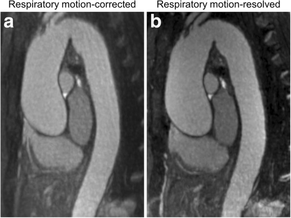
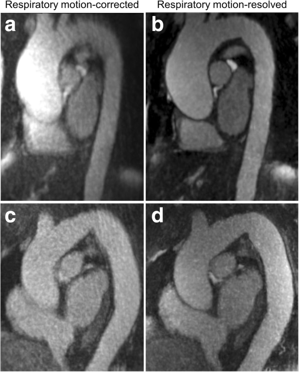
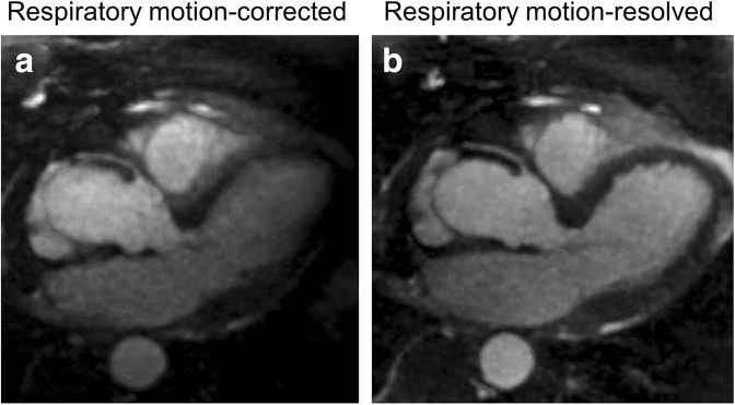
Background: Whole-heart magnetic resonance angiography (MRA) requires sophisticated methods accounting for respiratory motion. Our purpose was to evaluate the image quality of compressed sensing-based respiratory motion-resolved three-dimensional (3D) whole-heart MRA compared with self-navigated motion-corrected whole-heart MRA in patients with known thoracic aorta dilation.
Methods: Twenty-five patients were prospectively enrolled in this ethically approved study. Whole-heart 1.5-T MRA was acquired using a prototype 3D radial steady-state free-precession free-breathing sequence. The same data were reconstructed with a one-dimensional motion-correction algorithm (1D-MCA) and an extradimensional golden-angle radial sparse parallel reconstruction (XD-GRASP). Subjective image quality was scored and objective image quality was quantified (signal intensity ratio, SIR; vessel sharpness). Wilcoxon, McNemar, and paired t tests were used.
Results: Subjective image quality was significantly higher using XD-GRASP compared to 1D-MCA (median 4.5, interquartile range 4.5-5.0 versus 4.0 [2.25-4.75]; p < 0.001), as well as signal homogeneity (3.0 [3.0-3.0] versus 2.0 [2.0-3.0]; p = 0.003), and image sharpness (3.0 [2.0-3.0] vs 2.0 [1.25-3.0]; p < 0.001). SIR with the 1D-MCA and XD-GRASP was 6.1 ± 3.9 versus 7.4 ± 2.5, respectively (p < 0.001); while signal homogeneity was 274.2 ± 265.0 versus 199.8 ± 67.2 (p = 0.129). XD-GRASP provided a higher vessel sharpness (45.3 ± 10.7 versus 40.6 ± 101, p = 0.025).
Conclusions: XD-GRASP-based motion-resolved reconstruction of free-breathing 3D whole-heart MRA datasets provides improved image contrast, sharpness, and signal homogeneity and seems to be a promising technique that overcomes some of the limitations of motion correction or respiratory navigator gating.
Keywords: Aorta; Dilatation; Image processing (computer–assisted); Magnetic resonance angiography; Motion.
Conflict of interest statement
U. Joseph Schoepf is a consultant for and/or receives research support from Astellas, Bayer, Elucid Bioimaging, GE, Guerbet, HeartFlow Inc., and Siemens. Davide Piccini is an employee of Siemens. John Heerfordt’s doctoral studies are financially supported by Siemens. Akos Varga-Szemes receives institutional research support and travel support from Siemens and is a consultant for Elucid Bioimaging. The other authors declare that they have no competing interests.
Figures
Similar articles
-
Motion compensated whole-heart coronary cardiovascular magnetic resonance angiography using focused navigation (fNAV).J Cardiovasc Magn Reson. 2021 Mar 29;23(1):33. doi: 10.1186/s12968-021-00717-4. J Cardiovasc Magn Reson. 2021. PMID: 33775246 Free PMC article.
-
Optimized respiratory-resolved motion-compensated 3D Cartesian coronary MR angiography.Magn Reson Med. 2018 Dec;80(6):2618-2629. doi: 10.1002/mrm.27208. Epub 2018 Apr 22. Magn Reson Med. 2018. PMID: 29682783 Free PMC article.
-
Four-dimensional respiratory motion-resolved whole heart coronary MR angiography.Magn Reson Med. 2017 Apr;77(4):1473-1484. doi: 10.1002/mrm.26221. Epub 2016 Mar 28. Magn Reson Med. 2017. PMID: 27052418 Free PMC article.
-
Respiratory Motion-Resolved Compressed Sensing Reconstruction of Free-Breathing Radial Acquisition for Dynamic Liver Magnetic Resonance Imaging.Invest Radiol. 2015 Nov;50(11):749-56. doi: 10.1097/RLI.0000000000000179. Invest Radiol. 2015. PMID: 26146869 Free PMC article.
-
High efficiency free-breathing 3D thoracic aorta vessel wall imaging using self-gating image reconstruction.Magn Reson Imaging. 2024 Apr;107:80-87. doi: 10.1016/j.mri.2024.01.009. Epub 2024 Jan 17. Magn Reson Imaging. 2024. PMID: 38237694 Review.
Cited by
-
Measurement accuracy of prototype non-contrast, compressed sensing-based, respiratory motion-resolved whole heart cardiovascular magnetic resonance angiography for the assessment of thoracic aortic dilatation: comparison with computed tomography angiography.J Cardiovasc Magn Reson. 2021 Feb 8;23(1):7. doi: 10.1186/s12968-020-00697-x. J Cardiovasc Magn Reson. 2021. PMID: 33557887 Free PMC article.
-
Latest Advances in Image Acceleration: All Dimensions are Fair Game.J Magn Reson Imaging. 2023 Feb;57(2):387-402. doi: 10.1002/jmri.28462. Epub 2022 Oct 7. J Magn Reson Imaging. 2023. PMID: 36205716 Free PMC article. Review.
-
Motion compensated whole-heart coronary cardiovascular magnetic resonance angiography using focused navigation (fNAV).J Cardiovasc Magn Reson. 2021 Mar 29;23(1):33. doi: 10.1186/s12968-021-00717-4. J Cardiovasc Magn Reson. 2021. PMID: 33775246 Free PMC article.
-
Deep learning-based left ventricular segmentation demonstrates improved performance on respiratory motion-resolved whole-heart reconstructions.Front Radiol. 2023 Jun 2;3:1144004. doi: 10.3389/fradi.2023.1144004. eCollection 2023. Front Radiol. 2023. PMID: 37492382 Free PMC article.
-
Accelerating 3D MTC-BOOST in patients with congenital heart disease using a joint multi-scale variational neural network reconstruction.Magn Reson Imaging. 2022 Oct;92:120-132. doi: 10.1016/j.mri.2022.06.012. Epub 2022 Jun 27. Magn Reson Imaging. 2022. PMID: 35772584 Free PMC article.
References
Publication types
MeSH terms
LinkOut - more resources
Full Text Sources