Comment
. 2019 Aug 15;38(16):e102799.
doi: 10.15252/embj.2019102799. Epub 2019 Jul 31.

Antigen cross-presentation: proteasome location, location, location

Affiliations
Comment

Antigen cross-presentation: proteasome location, location, location

Michel Desjardins. EMBO J. .

Abstract

Our understanding of the mechanisms by which peptides from proteins present in phagosomes and endosomes are processed and presented on MHC class I molecules, in a pathway called cross-presentation, is still incomplete. One of the main questions arising from currently proposed models is how do proteins in the phagosome lumen reach the proteasome in the cytoplasm to be processed properly. In this issue of The EMBO Journal, Sengupta et al (2019) present evidence for a surprising turn of events where, in fact, the proteasome acts within the lumen of endosomes and phagosomes.


Figures

Figure 1
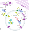
Figure 1. Models for the location of the proteasome in cross‐presentation
The proteasome can be positioned in various locations within the cell to accommodate the need to process large polypeptides into the smaller peptides that fit best on the groove of MHC I molecules. (1) In the phagosome‐to‐cytosol pathway, large polypeptides are translocated from the lumen into the cytoplasm by an unknown transporter to reach the proteasome. Antigenic peptides enter the ER lumen through the TAP transporters of the classical MHC I pathway. (2) The proteasome is closely associated with the phagosome membrane (in the cytoplasmic side). Large polypeptides translocated as in 1 reach the proteasome and the peptides are moved back into the phagosome lumen through TAP to bind MHC I molecules. (3) The positioning of the proteasome in the phagosome lumen has the advantage to enable a very efficient system to generate peptides binding directly to MHC I molecules in the lumen (acquired through ER–phagosome interaction). This processing pathway, which is more efficient than the potential processing by hydrolases, would take place in a TAP‐independent process.

Comment on

References

    1. Ackerman AL, Kyritsis C, Tampe R, Cresswell P (2003) Early phagosomes in dendritic cells form a cellular compartment sufficient for cross presentation of exogenous antigens. Proc Natl Acad Sci USA 100: 12889–12894 - PMC - PubMed
    1. Bevan MJ (1976) Cross‐priming for a secondary cytotoxic response to minor H antigens with H‐2 congenic cells which do not cross‐react in the cytotoxic assay. J Exp Med 143: 1283–1288 - PMC - PubMed
    1. English L, Chemali M, Duron J, Rondeau C, Laplante A, Gingras D, Alexander D, Leib D, Norbury C, Lippe R et al (2009) Autophagy enhances the presentation of endogenous viral antigens on MHC class I molecules during HSV‐1 infection. Nat Immunol 10: 480–487 - PMC - PubMed
    1. Gagnon E, Duclos S, Rondeau C, Chevet E, Cameron PH, Steele‐Mortimer O, Paiement J, Bergeron JJ, Desjardins M (2002) Endoplasmic reticulum‐mediated phagocytosis is a mechanism of entry into macrophages. Cell 110: 119–131 - PubMed
    1. Guermonprez P, Saveanu L, Kleijmeer M, Davoust J, Van Endert P, Amigorena S (2003) ER‐phagosome fusion defines an MHC class I cross‐presentation compartment in dendritic cells. Nature 425: 397–402 - PubMed

Substances