. 2019 Oct 1;317(4):F815-F824.
doi: 10.1152/ajprenal.00196.2019. Epub 2019 Jul 31.

Cellular cholesterol modifies flow-mediated gene expression

Affiliations

Cellular cholesterol modifies flow-mediated gene expression

Robert L Repetti et al. Am J Physiol Renal Physiol. .

Abstract

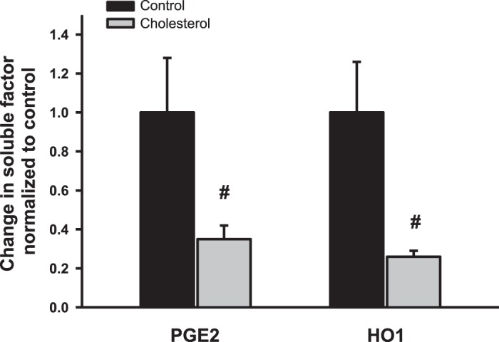
Downregulation of heme oxygenase-1 (HO-1), cyclooxygenase-2 (COX2), and nitric oxide synthase-2 (NOS2) in the kidneys of Dahl rodents causes salt sensitivity, while restoring their expression aids in Na+ excretion and blood pressure reduction. Loading cholesterol into collecting duct (CD) cells represses fluid shear stress (FSS)-mediated COX2 activity. Thus, we hypothesized that cholesterol represses flow-responsive genes necessary to effectuate Na+ excretion. To this end, CD cells were used to test whether FSS induces these genes and if cholesterol loading represses them. Mice fed either 0% or 1% cholesterol diet were injected with saline, urine volume and electrolytes were measured, and renal gene expression determined. FSS-exposed CD cells demonstrated increases in HO-1 mRNA by 350-fold, COX2 by 25-fold, and NOS2 by 8-fold in sheared cells compared with static cells (P < 0.01). Immunoblot analysis of sheared cells showed increases in HO-1, COX2, and NOS2 protein, whereas conditioned media contained more HO-1 and PGE2 than static cells. Cholesterol loading repressed the sheared mediated protein abundance of HO-1 and NOS2 as well as HO-1 and PGE2 concentrations in media. In cholesterol-fed mice, urine volume was less at 6 h after injection of isotonic saline (P < 0.05). Urinary Na+ concentration, urinary K+ concentration, and osmolality were greater, whereas Na+ excretion was less, at the 6-h urine collection time point in cholesterol-fed versus control mice (P < 0.05). Renal cortical and medullary HO-1 (P < 0.05) and NOS2 (P < 0.05) mRNA were repressed in cholesterol-fed compared with control mice. Cholesterol acts to repress flow induced natriuretic gene expression, and this effect, in vivo, may contribute to renal Na+ avidity.

Keywords: blood pressure; collecting duct; flow; shear.


Conflict of interest statement

No conflicts of interest, financial or otherwise, are declared by the author(s).

Figures

Fig. 1.
Fig. 1.
Fluid shear stress (FSS) induces heme oxygenase-1 (HO1), cyclooxygenase-2 (COX2), and nitric oxide synthase-2 (NOS2) mRNA expression compared with static controls. Inner medullary collecting duct 3 cells were exposed to 0.4 dyn/cm2 of FSS (n = 4–6) or maintained under static conditions (n = 3–6) from 2 to 6 h. Total RNA was extracted from shear-exposed and static cells, and PCR was performed using specific gene-specific TaqMan primers. Steady-state mRNA expression of HO-1 (51.8 ± 6.2-, 63.0 ± 2.6-, and 351 ± 35-fold increase, respectively) and COX2 (6.3 ± 0.7-, 4.0 ± 0.7-, and 29.5 ± 2.0-fold increase, respectively) increased at 2, 4, and 6 h in shear-exposed cells versus static control cells (#P < 0.05). NOS2 mRNA abundance increased above static control at 4 and 6 h (2.5 ± 0.2- and 8.5 ± 0.9-fold, respectively; #P < 0.05 vs. the static group).
Fig. 2.
Fig. 2.
Protein abundance of cyclooxygenase-2 (COX2) and heme oxygenase-1 (HO-1) in inner medullary collecting duct 3 (IMCD3) cells exposed for up to 4 h. A: COX2 and HO-1 protein expression by Western blot analysis in fluid shear stress (FSS)-exposed and static IMCD3 cells. B: densitometry demonstrating no difference in COX2 expression at 2 h; however, at 4 h, shear-exposed cells expressed ~2.5-fold increased COX2 (#P < 0.05) compared with static cells. C: HO-1 densitometry similarly showed no effect of FSS at 2 h; however, at 4 h, there was an ~6-fold (#P < 0.05) increase in protein abundance compared with static cells.
Fig. 3.
Fig. 3.
Nitric oxide synthase-2 (NOS2) protein expression increases at 2 and 4 h in fluid shear stress (FSS)-exposed cells. A: Western blot analysis of NOS2 expression in cells exposed to 0.4 dyn/cm2 of FSS versus static controls. B: NOS2 abundance was greater in shear-exposed cells at 2 h (#P < 0.05) and 4 h (#P < 0.05) versus static controls.
Fig. 4.
Fig. 4.
Fluid shear stress (FSS) induces cyclooxygenase-2 (COX2), nitric oxide synthase-2 (NOS2), and heme oxygenase-1 (HO-1) abundance at 6 h. A: Western blot analysis of protein lysates generated from inner medullary collecting duct 3 cells exposed to FSS versus static controls to measure immunodetectable COX2, NOS2, and HO-1. B: densitometry illustrating the ~2-fold increase in COX2, ~6-fold increase in NOS2, and ~20-fold increase in HO-1 in FSS-exposed cells (#P < 0.05) compared with static cells.
Fig. 5.
Fig. 5.
PGE2 and heme oxygenase-1 (HO-1) expression in the media of fluid shear stress (FSS)-exposed and static cells. A: PGE2 concentration in the media bathing the cells, normalized to the total amount of protein lysate, was significantly greater at each time point compared with static controls (#P < 0.05). B: HO-1 concentration in the media bathing the cells, normalized to the total amount of protein lysate, was not different at 2 h but was significantly greater at 4 and 6 h compared with static controls (#P < 0.05).
Fig. 6.
Fig. 6.
Cellular cholesterol content after cholesterol loading and this effect on ATP-binding cassette subfamily A member 1 (ABCA1) gene expression. A: inner medullary collecting duct 3 (IMCD3) cells were loaded with a cholesterol/methyl-β-cyclodextrin mixture for ~36 h, whereas control cells were maintained under serum-free conditions. Cellular cholesterol content was ~2-fold greater in cholesterol-loaded cells (#P < 0.05) versus control cells. B: ABCA1 expression was increased >3-fold in cholesterol-treated cells (#P < 0.05) compared with control cells.
Fig. 7.
Fig. 7.
Cholesterol-loaded cells express less shear-induced nitric oxide synthase-2 (NOS2) and heme oxygenase-1 (HO-1) than control cells after 4 h of fluid shear stress (FSS). A: Western blot analysis of control and cholesterol-treated inner medullary collecting duct 3 cells after exposure to 4 h of FSS. Steady-state protein abundance of NOS2 and HO-1 was less in cholesterol-treated versus control cells. B: densitometric analysis of control cells (n = 22–24) and cholesterol-loaded cells (n = 22–24) cells confirmed that NOS2 and HO-1 protein expression was reduced in cholesterol-treated cells (*P < 0.05 vs. control cells); however, COX2 was unchanged.
Fig. 8.
Fig. 8.
Soluble PGE2 and heme oxygenase-1 (HO-1) are reduced in conditioned media of cholesterol-treated compared with control inner medullary collecting duct 3 cells after 4 h of fluid shear stress. Concentrations of PGE2 and HO-1 were determined (in pg/ml) and normalized to the quantity of cellular protein lysate (in µg). Data sets were then normalized to the control shear. PGE2 and HO-1 in conditioned media was only 0.35 ± 0.07 and 0.26 ± 0.03 in cholesterol-loaded cells (#P < 0.05), respectively, compared with control sheared cells.
Fig. 9.
Fig. 9.
Urine volume as a percentage of injected saline measured sequentially from 2 to 6 h in control and cholesterol-fed mice. Male FVB mice fed either 0% or 1% cholesterol diet for 6 wk were placed into a metabolic cage and injected with isotonic saline at 5% of body weight, and urine was collected sequentially from 2 to 6 h. Urine volume is presented as a percentage of the injected isotonic saline by volume. No difference in excretion of the saline load was noted between 2 and 4 h; however, at 6 h, urine volume as a percentage of injected saline in the cholesterol-fed group was less than 50% of the control group. No difference was noted between total urine volumes excreted over 6 h.
Fig. 10.
Fig. 10.
Renal cortical (A) and medullary (B) mRNA expression of heme oxygenase-1 (HO-1), cyclooxygenase-2 (COX2), and nitric oxide synthase-2 (NOS2) in 6- to 8-wk control and cholesterol-fed mice. After injection of isotonic saline and urine collection, control and cholesterol-fed mice were euthanized, the renal cortex and medulla was extracted, and total RNA was generated. Steady-state abundances of HO-1 and NOS2 were reduced in the cortex (A) and medulla (B) of cholesterol-fed mice (#P < 0.05) versus control mice, whereas no difference was noted in COX2 expression.
Fig. 11.
Fig. 11.
Renal immunoblot expression of cyclooxygenase-2 (COX2) did not differ between control and cholesterol-fed mice. Total protein lysates from kidneys of mice fed 0% (n = 8) and 1% (n = 8) cholesterol diet for 6–8 wk were immunoblotted for COX2 and β-actin. Although no statistical difference in expression was noted between control and cholesterol-fed mice, steady-state renal medullary expression of COX2 tended (*P = 0.08) to be less in cholesterol-fed mice.
Fig. 12.
Fig. 12.
Renal immunoblot expression of nitric oxide synthase-2 (NOS2) was less in cholesterol-fed compared with control mice. Total protein lysates from kidneys of mice fed 0% (n = 8) and 1% (n = 8) cholesterol diet for 6–8 wk were immunoblotted for NOS2 and β-actin. NOS2 tended to be less in the renal cortex (*P = 0.08) of cholesterol-fed versus control mice, whereas NOS2 in the renal medulla of cholesterol-fed mice was significantly less (#P = 0.05) compared with control mice.
Fig. 13.
Fig. 13.
Renal immunoblot expression of heme oxygenase-1 (HO-1) did not differ between control and cholesterol-fed mice. Total protein lysates from kidneys of mice fed 0% (n = 4) and 1% (n = 4) cholesterol diet for 6–8 wk were immunoblotted for HO-1 and β-actin. Although no statistical difference in expression was noted between control and cholesterol-fed mice, steady-state renal medullary expression of HO-1 tended (*P = 0.06) to be less in cholesterol-fed mice.

Comment in

  • Cholesterol may not have a special place in kidneys.
    Nauli SM. Nauli SM. Am J Physiol Renal Physiol. 2019 Nov 1;317(5):F1169-F1170. doi: 10.1152/ajprenal.00394.2019. Epub 2019 Sep 18. Am J Physiol Renal Physiol. 2019. PMID: 31532244 Free PMC article. No abstract available.

References

    1. Althaus M, Fronius M, Buchäckert Y, Vadász I, Clauss WG, Seeger W, Motterlini R, Morty RE. Carbon monoxide rapidly impairs alveolar fluid clearance by inhibiting epithelial sodium channels. Am J Respir Cell Mol Biol 41: 639–650, 2009. doi:10.1165/rcmb.2008-0458OC. - DOI - PubMed
    1. Awayda MS, Awayda KL, Pochynyuk O, Bugaj V, Stockand JD, Ortiz RM. Acute cholesterol-induced anti-natriuretic effects: role of epithelial Na+ channel activity, protein levels, and processing. J Biol Chem 286: 1683–1695, 2011. doi:10.1074/jbc.M110.159194. - DOI - PMC - PubMed
    1. Bugaj V, Pochynyuk O, Mironova E, Vandewalle A, Medina JL, Stockand JD. Regulation of the epithelial Na+ channel by endothelin-1 in rat collecting duct. Am J Physiol Renal Physiol 295: F1063–F1070, 2008. doi:10.1152/ajprenal.90321.2008. - DOI - PMC - PubMed
    1. Cai Z, Xin J, Pollock DM, Pollock JS. Shear stress-mediated NO production in inner medullary collecting duct cells. Am J Physiol Renal Physiol 279: F270–F274, 2000. doi:10.1152/ajprenal.2000.279.2.F270. - DOI - PubMed
    1. Flores D, Battini L, Gusella GL, Rohatgi R. Fluid shear stress induces renal epithelial gene expression through polycystin-2-dependent trafficking of extracellular regulated kinase. Nephron Physiol 117: 27–36, 2011. doi:10.1159/000321640. - DOI - PMC - PubMed

Publication types

LinkOut - more resources