. 2019 Oct 1;317(4):F922-F929.
doi: 10.1152/ajprenal.00375.2018. Epub 2019 Jul 31.

Cell-free hemoglobin augments acute kidney injury during experimental sepsis

Affiliations

Cell-free hemoglobin augments acute kidney injury during experimental sepsis

Ciara M Shaver et al. Am J Physiol Renal Physiol. .

Abstract

Acute kidney injury is a common complication of severe sepsis and contributes to high mortality. The molecular mechanisms of acute kidney injury during sepsis are not fully understood. Because hemoproteins, including myoglobin and hemoglobin, are known to mediate kidney injury during rhabdomyolysis, we hypothesized that cell-free hemoglobin (CFH) would exacerbate acute kidney injury during sepsis. Sepsis was induced in mice by intraperitoneal injection of cecal slurry (CS). To mimic elevated levels of CFH observed during human sepsis, mice also received a retroorbital injection of CFH or dextrose control. Four groups of mice were analyzed: sham treated (sham), CFH alone, CS alone, and CS + CFH. The addition of CFH to CS reduced 48-h survival compared with CS alone (67% vs. 97%, P = 0.001) and increased the severity of illness. After 24 and 48 h, CS + CFH mice had a reduced glomerular filtration rate from baseline, whereas sham, CFH, and CS mice maintained baseline glomerular filtration rate. Biomarkers of acute kidney injury, neutrophil gelatinase-associated lipocalin (NGAL) and kidney injury molecule-1 (KIM-1), were markedly elevated in CS+CFH compared with CS (8-fold for NGAL and 2.4-fold for KIM-1, P < 0.002 for each) after 48 h. Histological examination showed a trend toward increased tubular injury in CS + CFH-exposed kidneys compared with CS-exposed kidneys. However, there were similar levels of renal oxidative injury and apoptosis in the CS + CFH group compared with the CS group. Kidney levels of multiple proinflammatory cytokines were similar between CS and CS + CFH groups. Human renal tubule cells (HK-2) exposed to CFH demonstrated increased cytotoxicity. Together, these results show that CFH exacerbates acute kidney injury in a mouse model of experimental sepsis, potentially through increased renal tubular injury.

Keywords: acute kidney injury; cell-free hemoglobin; sepsis.


Conflict of interest statement

No conflicts of interest, financial or otherwise, are declared by the authors.

Figures

Fig. 1.
Fig. 1.
Cell-free hemoglobin (CFH) reduces survival during experimental sepsis. BALB/c mice were given intraperitoneal injection of cecal slurry (CS) or 5% dextrose followed by retroorbital injection of CFH or PBS. CS + CFH mice had reduced survival compared with CFH alone, sham-treated (sham), and CS mice (*P = 0.001, linear-by-linear association). n = 15 mice in the sham group, 4 mice in the CFH group, 29 mice in the CS group, and 26 mice in the CS + CFH group.
Fig. 2.
Fig. 2.
Cell-free hemoglobin (CFH) increases the severity of illness without affecting weight loss during experimental sepsis. A: CFH during sepsis (CS + CFH) increased the severity of illness compared with cecal slurry (CS), sham-treated (sham), or CFH mice as measured by a composite sepsis score. B: CS and CS + CFH mice had increased weight loss compared with sham mice (P < 0.001). n = 4–19 mice/group.
Fig. 3.
Fig. 3.
Cell-free hemoglobin (CFH) reduced glomerular filtration rate (GFR) during experimental sepsis. GFR was measured at baseline and after 24 and 48 h of experimental sepsis and adjusted for body weight at the time of assessment. There was no change in GFR over time in sham-treated (sham), CFH, or cecal slurry (CS)-treated mice. GFR significantly declined in mice exposed to CS + CFH (*P = 0.013 and **P = 0.008 by a Wilcoxon signed-rank test). n = 6–13 mice/group.
Fig. 4.
Fig. 4.
Cell-free hemoglobin (CFH) augments expression of acute kidney injury biomarkers. A: expression of the acute kidney injury biomarker neutrophil gelatinase-associated lipocalin (NGAL) was increased in whole kidney homogenates from mice with sepsis [cecal slurry (CS)] and was augmented by CFH (CS + CFH). B: expression of the acute kidney injury biomarker kidney injury molecule-1 (Kim-1) was increased in whole kidney homogenates from mice with sepsis (CS) and was augmented by cell-free hemoglobin (CS + CFH). n = 4–24 mice/group.
Fig. 5.
Fig. 5.
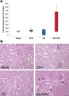
Cell-free hemoglobin (CFH) increases renal tubular injury in the setting of sepsis. A: quantification of tubular injury scores showed a trend toward increased injury in cecal slurry (CS) + CFH (P = 0.353 for Kruskal-Wallis comparisons and P = 0.137 for CS vs. CS + CFH comparison by a Mann-Whitney U-test). Data were normalized to percent sham treatment (sham) values because of day-to-day variability in the degree of renal injury present in control animals. B: representative images of periodic acid Schiff-stained kidney sections from each experimental group. Arrows indicate areas of irregular-appearing detached tubular epithelial cells. *Peritubular capillary expansion containing eosinophilic material. n = 5–9 per group.
Fig. 6.
Fig. 6.
The effect of cell-free hemoglobin (CFH) in the kidney is not due to oxidative stress or apoptosis. A–C: levels of F2-isoprostanes (A; P = 0.883), isofurans (B; P = 0.563), and malondialdehyde (MDA; C; P = 0.868) were similar in kidneys of mice treated with cecal slurry (CS) or CS with CFH (CS + CFH). D: there was no evidence of increased apoptosis in kidneys of septic mice (P = 0.442), although CFH in the absence of sepsis may result in apoptosis. n = 6–15 per group. HPF, high-powered field; sham, sham treatment.
Fig. 7.
Fig. 7.
Cell-free hemoglobin increases cytotoxicity of tubular epithelial cells in vitro. HK-2 cells were exposed to increasing concentrations of cell-free hemoglobin (0–1 mg/ml) for 24 h, and viability was assessed by lactate dehydrogenase release. Statistical significance was measured by Student’s t-tests compared with control. n = 3 per group.

References

    1. Adamzik M, Hamburger T, Petrat F, Peters J, de Groot H, Hartmann M. Free hemoglobin concentration in severe sepsis: methods of measurement and prediction of outcome. Crit Care 16: R125, 2012. doi:10.1186/cc11425. - DOI - PMC - PubMed
    1. Alobaidi R, Basu RK, Goldstein SL, Bagshaw SM. Sepsis-associated acute kidney injury. Semin Nephrol 35: 2–11, 2015. doi:10.1016/j.semnephrol.2015.01.002. - DOI - PMC - PubMed
    1. Baek JH, D’Agnillo F, Vallelian F, Pereira CP, Williams MC, Jia Y, Schaer DJ, Buehler PW. Hemoglobin-driven pathophysiology is an in vivo consequence of the red blood cell storage lesion that can be attenuated in guinea pigs by haptoglobin therapy. J Clin Invest 122: 1444–1458, 2012. doi:10.1172/JCI59770. - DOI - PMC - PubMed
    1. Bastarache JA, Koyama T, Wickersham NE, Ware LB. Validation of a multiplex electrochemiluminescent immunoassay platform in human and mouse samples. J Immunol Methods 408: 13–23, 2014. doi:10.1016/j.jim.2014.04.006. - DOI - PMC - PubMed
    1. Bellomo R, Kellum JA, Ronco C. Acute kidney injury. Lancet 380: 756–766, 2012. doi:10.1016/S0140-6736(11)61454-2. - DOI - PubMed

Publication types

MeSH terms