. 2020 Mar 14;30(3):1001-1015.
doi: 10.1093/cercor/bhz143.

Uncovering a Role for the Dorsal Hippocampal Commissure in Recognition Memory

Affiliations

Uncovering a Role for the Dorsal Hippocampal Commissure in Recognition Memory

M Postans et al. Cereb Cortex. .

Abstract

The dorsal hippocampal commissure (DHC) is a white matter tract that provides interhemispheric connections between temporal lobe brain regions. Despite the importance of these regions for learning and memory, there is scant evidence of a role for the DHC in successful memory performance. We used diffusion-weighted magnetic resonance imaging (DW-MRI) and white matter tractography to reconstruct the DHC in both humans (in vivo) and nonhuman primates (ex vivo). Across species, our findings demonstrate a close consistency between the known anatomy and tract reconstructions of the DHC. Anterograde tract-tracer techniques also highlighted the parahippocampal origins of DHC fibers in nonhuman primates. Finally, we derived diffusion tensor MRI metrics from the DHC in a large sample of human subjects to investigate whether interindividual variation in DHC microstructure is predictive of memory performance. The mean diffusivity of the DHC correlated with performance in a standardized recognition memory task, an effect that was not reproduced in a comparison commissure tract-the anterior commissure. These findings highlight a potential role for the DHC in recognition memory, and our tract reconstruction approach has the potential to generate further novel insights into the role of this previously understudied white matter tract in both health and disease.

Keywords: familiarity; hippocampal commissure; recognition memory; recollection; tractography.


Figures

Figure 1
Figure 1
ROIs used for DHC and AC tractography. The hippocampal (blue) and manually drawn (red lines) ROIs used for DHC tractography shown on a midsagittal section of a T1-weighted image for a representative ex vivo nonhuman primate specimen in a 0.5-mm3 native diffusion space (A) and an HCP subject in a 1.25-mm3 native diffusion space (B). Also shown are the manually-drawn ROIs used for AC tractography (red lines and blue square) in a representative HCP subject (C).
Figure 2
Figure 2
ROIs used to extract the DHC and the subsequent tract reconstructions. (A) The hippocampal (blue) and manually-drawn (red lines) ROIs used to extract the DHC in one representative specimen and the subsequent reconstructions shown over a midsagittal and coronal section from the corresponding T1-weighted image in a 0.5-mm3 native diffusion space. (B) The reconstructions in all 4 specimens. The DHC reconstructions illustrated from a left-lateral, anterior–posterior, and ventral perspective, alongside the anatomical hippocampal ROIs for spatial context (C, D, and E, respectively). Note that, for computational purposes, these renderings contain a one-eighth subsample of all reconstructed streamlines.
Figure 3
Figure 3
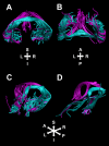
DHC (teal) and fornix (purple) streamlines reconstructed in representative cases. Streamlines corresponding to these tracts are shown for a representative ex vivo nonhuman primate specimen, shown from rear coronal oblique (A), ventral (B), and left-lateral oblique (C) perspectives. For comparison, streamlines corresponding to these 2 tracts are also shown for a representative HCP subject, from a left-lateral oblique perspective (D).
Figure 4
Figure 4
DHC (teal), tapetum (yellow), and fornix (purple) streamlines reconstructed in representative cases. (A) Streamlines corresponding to the DHC and tapetum in a nonhuman primate case (left) and an HCP subject (right), against a midsagittal T1-weighted image section. (B, C) The same streamlines from anterior-coronal and ventral perspectives, respectively. (D, E) The same streamlines alongside those corresponding to the fornix.
Figure 5
Figure 5
Bright- and dark-field photomicrographs of coronal sections taken at the level of the DHC, inferior to the corpus callosum. A1 contains a bright-field photomicrograph from case ACy28, which received a large tracer injection that filled much of the caudal hippocampus, including the subiculum. Dense anterograde label can be seen in the medial fornix alongside a lighter label seemingly entering the DHC at the level of the inferior forceps. The large majority of this label then turned rostral to occupy the medial half of the DHC, as seen in A2. A2 is from the same case but more anterior (at the level of the habenula). Some of the label in the DHC decussates to join the medial contralateral fornix. (B, C) Dark-field photomicrographs from case ACy14 (coronal section at the level of the splenium), which received a tracer injection centered in the rostral subiculum. A very clear label is present in the ipsilateral (B), but not contralateral (C), fornix, while no label is apparent in the DHC at the level of the inferior forceps. (D, E) Dark-field photomicrographs from case ACyF23, whose injection incorporated the caudal perirhinal and anterior parahippocampal cortices. Light label was evident in the most medial fornix, alongside labeled fibers in both the left and right DHC, within the inferior forceps. Magnified inserts are included in panels A1, A2, D, and E. All scale bars = 200 μm. Abbreviations: CC, corpus callosum; F, fornix; H, habenula; if, inferior forceps; Rspl, retrosplenial cortex.
Figure 6
Figure 6
The DHC reconstructions in 4 representative HCP datasets and structure–cognition correlations reported in the text. DHC reconstructions are shown in the coronal (A) and axial (B) planes. The correlations between white matter MD and CPWM total scores in the DHC and AC are illustrated (in C and D, respectively). The best-fitting linear regression line is plotted alongside 95% CIs.

References

    1. Aggleton JP. 2008. EPS Mid-Career Award 2006. Understanding anterograde amnesia: disconnections and hidden lesions. Q J Exp Psychol (Hove). 61:1441–1471. - PubMed
    1. Aggleton JP. 2012. Multiple anatomical systems embedded within the primate medial temporal lobe: implications for hippocampal function. Neurosci Biobehav Rev. 36:1579–1596. - PubMed
    1. Aggleton JP, Brown MW. 1999. Episodic memory, amnesia, and the hippocampal-anterior thalamic axis. Behav Brain Sci. 22:425–444discussion 444–489. - PubMed
    1. Aggleton JP, Desimone R, Mishkin M. 1986. The origin, course, and termination of the hippocampothalamic projections in the macaque. J Comp Neurol. 243:409–421. - PubMed
    1. Amaral RSC, Park MTM, Devenyi GA, Lynn V, Pipitone J, Winterburn J, Chavez S, Schira M, Lobaugh NJ, Voineskos AN et al. . 2018. Manual segmentation of the fornix, fimbria, and alveus on high-resolution 3T MRI: application via fully-automated mapping of the human memory circuit white and grey matter in healthy and pathological aging. Neuroimage. 170:132–150. - PubMed

Publication types