. 2019 Nov;49(11):1487-1499.
doi: 10.1111/cea.13475. Epub 2019 Aug 26.

Syngeneic red blood cell-induced extracellular vesicles suppress delayed-type hypersensitivity to self-antigens in mice

Affiliations

Syngeneic red blood cell-induced extracellular vesicles suppress delayed-type hypersensitivity to self-antigens in mice

Katarzyna Nazimek et al. Clin Exp Allergy. 2019 Nov.

Abstract

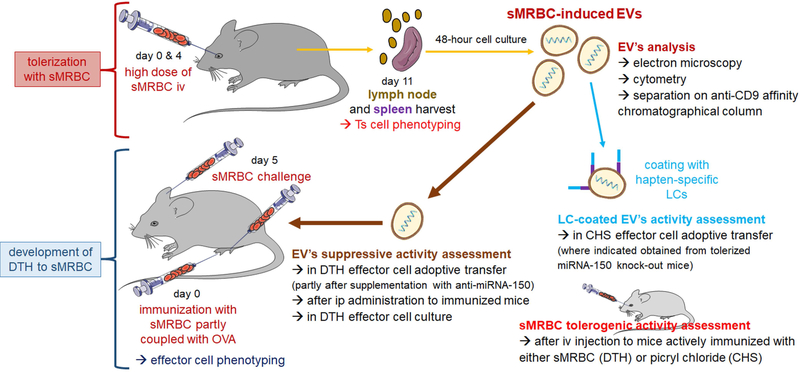
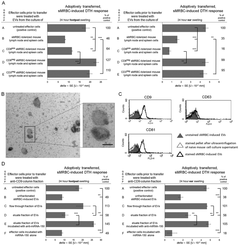
Background: At present, the role of autologous cells as antigen carriers inducing immune tolerance is appreciated. Accordingly, intravenous administration of haptenated syngeneic mouse red blood cells (sMRBC) leads to hapten-specific suppression of contact hypersensitivity (CHS) in mice, mediated by light chain-coated extracellular vesicles (EVs). Subsequent studies suggested that mice intravenously administered with sMRBC alone may also generate regulatory EVs, revealing the possible self-tolerogenic potential of autologous erythrocytes.

Objectives: The current study investigated the immune effects induced by mere intravenous administration of a high dose of sMRBC in mice.

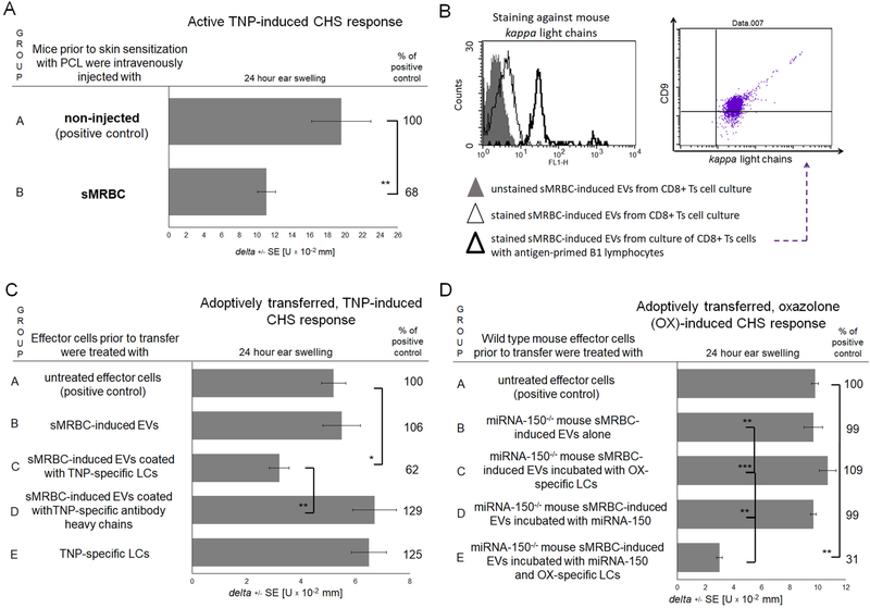
Methods: The self-tolerogenic potential of EVs was determined in a newly developed mouse model of delayed-type hypersensitivity (DTH) to sMRBC. The effects of EV's action on DTH effector cells were evaluated cytometrically. The suppressive activity of EVs, after coating with anti-hapten antibody light chains, was assessed in hapten-induced CHS in wild-type or miRNA-150-/- mice.

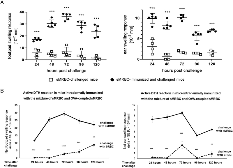
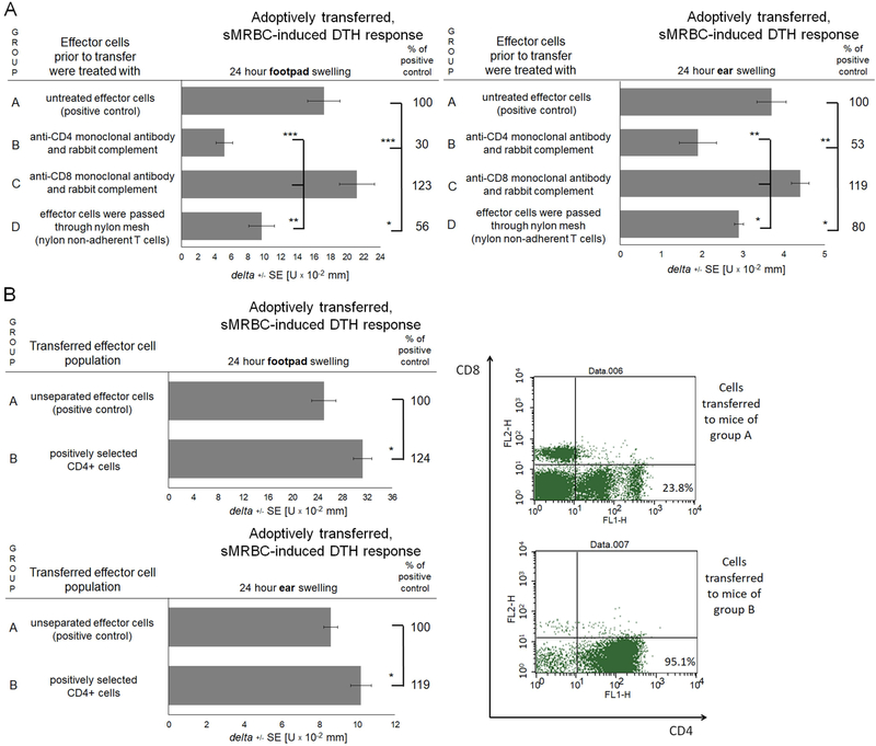
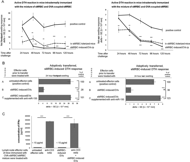
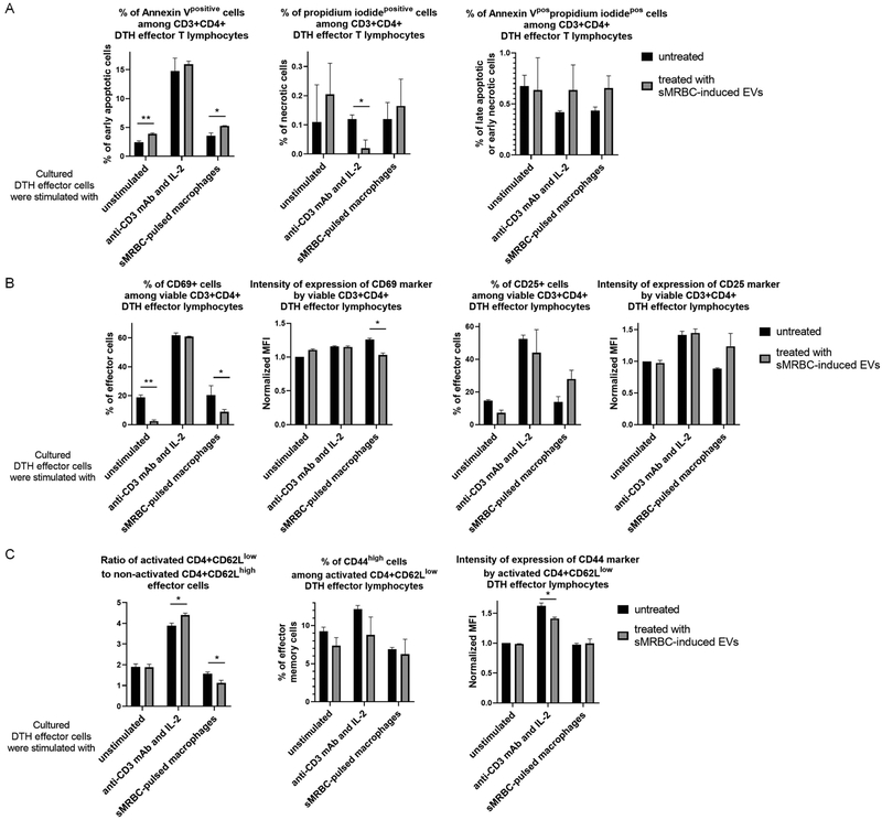
Results: Intravenous administration of sMRBC led to the generation of CD9 + CD81+ EVs that suppressed sMRBC-induced DTH in a miRNA-150-dependent manner. Furthermore, the treatment of DTH effector cells with sMRBC-induced EVs decreased the activation of T cells but enhanced their apoptosis. Finally, EVs coated with antibody light chains inhibited hapten-induced CHS.

Conclusions and clinical relevance: The current study describes a newly discovered mechanism of self-tolerance induced by the intravenous delivery of a high dose of sMRBC that is mediated by EVs in a miRNA-150-dependent manner. This mechanism implies the concept of naturally occurring immune tolerance, presumably activated by overloading of the organism with altered self-antigens.

Keywords: delayed-type hypersensitivity to self-antigen; extracellular vesicles; immune suppression; miRNA-150; mouse model of delayed-type hypersensitivity to self-antigen; self-tolerance.


Conflict of interest statement

Conflict of Interest

The Authors declare that they have no relevant conflicts of interest.

Figures

Fig. 1.
Fig. 1.. Experimental design.
To induce immune tolerance, mice were intravenously (iv) injected with a high dose of syngeneic red blood cells (sMRBC) at days 0 and 4, and 7 days later lymph nodes and spleens were collected for phenotyping and culturing of suppressor T (Ts) cells that release sMRBC-induced extracellular vesicles (EVs). Otherwise, mice were intradermally injected into footpads with sMRBC mixed with ovalbumin (OVA)-coupled sMRBC, and 5 days later were intradermally injected into footpads and ears with sMRBC alone to elicit delayed-type hypersensitivity (DTH) reaction, with phenotyping of effector cells. Further, sMRBC-induced EVs were analyzed for their biological properties and immune function in DTH and, after coating with hapten-specific antibody light chains (LCs), in contact hypersensitivity (CHS).
Fig. 2.
Fig. 2.. CBA mice can be antigen-specifically immunized with syngeneic mouse red blood cells (sMRBC) to elicit delayed-type hypersensitivity (DTH) reaction.
(A) Footpad (left graph) or ear (right graph) swelling response of mice that were either only challenged by intradermal injection of sMRBC (open squares) or firstly immunized by intradermal injection of sMRBC mixed with ovalbumin-coupled sMRBC into footpad skin and then similarly challenged by intradermal injection of sMRBC into footpad or ear skin (black dots), measured up to 120 hours after challenge. Bars represent the mean and standard deviation. (B) Footpad (left graph) or ear (right graph) swelling response of mice that were immunized by intradermal injection of sMRBC mixed with ovalbumin-coupled sMRBC into footpad skin and then challenged by intradermal injection of either sMRBC (solid line) or sheep red blood cells (SRBC, dotted line) into footpad or ear skin, measured up to 120 hours after challenge. Two-tailed Student’s T test (n=5). **p < 0.01, ***p < 0.005.
Fig. 3.
Fig. 3.. Delayed-type hypersensitivity (DTH) reaction to syngeneic mouse red blood cells (sMRBC) is mediated by lymph node and spleen CD4+ T cells.
(A) Footpad (left graph) or ear (right graph) swelling response of mouse recipients of either total population of lymph node and spleen DTH effector cells induced by intradermal injection of sMRBC mixed with ovalbumin-coupled sMRBC in group A or negatively selected DTH effector cells; i.e. depleted of CD4+ cells in group B, depleted of CD8+ cells in group C or deprived of nylon-adherent cells in group D. (B) Footpad (upper graph) or ear (lower graph) swelling response of mouse recipients of either total population of lymph node and spleen DTH effector cells induced by intradermal injection of sMRBC mixed with ovalbumin-coupled sMRBC in group A or positively selected CD4+ DTH effector cells in group B, with cytometric control of selection efficacy (right panel). One-way ANOVA with RIR Tukey test or two-tailed Student’s T test (n=5). *p < 0.05, **p < 0.01, ***p < 0.005.
Fig. 4.
Fig. 4.. Delayed-type hypersensitivity (DTH) reaction to syngeneic mouse red blood cells (sMRBC) can be suppressed by intravenous (iv) administration of sMRBC and the resulting extracellular vesicles (EVs) acting in a miRNA-150-dependent manner.
(A) Footpad (left graph) or ear (right graph) swelling response either of mice immunized by intradermal injection of sMRBC mixed with ovalbumin-coupled sMRBC (OVA-sMRBC) into footpad skin and then challenged by intradermal injection of sMRBC (solid line with circles), or of mice that had been intravenously administered with sMRBC prior to immunization (dotted line with circles), or of mice intraperitoneally (ip) administered with sMRBC-induced EVs 24 hours after challenge (solid line with squares), measured up to 120 hours after challenge. (B) Footpad (left graph) or ear (right graph) swelling response of mouse recipients of either untreated DTH effector cells induced by intradermal injection of sMRBC mixed with OVA-sMRBC in group A, or DTH effector cells treated prior to transfer with sMRBC-induced EVs in group B, or DTH effector cells treated prior to transfer with sMRBC-induced EVs supplemented with anti-miR-150 (miRNA-150 hairpin inhibitor) in group C. (C) The concentration of interferon gamma (IFNγ) released to 24-hour culture supernatant by lymph node DTH effector cells induced by intradermal injection of sMRBC mixed with OVA-sMRBC and cultured alone or in the presence of anti-CD3 monoclonal antibodies (mAb) and/or sMRBC-induced EVs. One-way ANOVA with RIR Tukey test (n=5). **p < 0.01, ***p < 0.005.
Fig. 5.
Fig. 5.. Syngeneic mouse red blood cell (sMRBC)-induced extracellular vesicles (EVs) are generated by CD3+CD8+ T cells and express CD9 and CD81 tetraspanins.
(A) Footpad (left graph) or ear (right graph) swelling response of mouse recipients of unaffected delayed-type hypersensitivity (DTH) effector cells in group A, or DTH effector cells pretreated with aMRBC-induced EVs produced by tolerized mouse lymph node and spleen cells cultured either as total population in group B, or after enrichment of CD8+ cells in group C, or after depleting of CD8+ or CD3+ cells in groups D and E, respectively. (B) Electron microscopy analysis of sMRBC-induced EVs (bar express 100 nm). (C) Cytometric analysis of sMRBC-induced EVs with staining against CD9, CD63 or CD81 vesicle markers. (D) Footpad (left graph) or ear (right graph) swelling response of mouse recipients of untreated DTH effector cells in group A, or DTH effector cells treated prior to transfer with either unfractionated sMRBC-induced EVs in group B, or EVs passing through anti-CD9 affinity column in group C, or EVs eluted from anti-CD9 column alone in group D or supplemented with anti-miR-150 (miRNA-150 hairpin inhibitor) in group E, or of recipients of DTH effector cells pretreated with miRNA-150 alone. One-way ANOVA with RIR Tukey test (n=4–5). *p < 0.05, **p < 0.01, ***p < 0.005.
Fig. 6.
Fig. 6.. Syngeneic mouse red blood cell (sMRBC)-induced extracellular vesicles (EVs) increase the apoptosis and decrease the activation of delayed-type hypersensitivity (DTH) effector cells.
(A) Cytometrically assessed percentage of CD3+CD4+ DTH effector cells that are positive in staining with annexin-V and/or propidium iodide, after 3-hour culture in the presence (gray bars) or absence (black bars) of sMRBC-induced EVs. (B) Percentage of viable CD3+CD4+ DTH effector cells that express either CD69 or CD25 markers and geometric mean of fluorescence intensity (MFI) of fluorescent dyes conjugated to monoclonal antibodies (mAb) binding to either CD69 or CD25 markers, cytometrically evaluated after 18- or 24-hour culture in the presence (gray bars) or absence (black bars) of sMRBC-induced EVs. (C) Ratio of activated to non-activated DTH effector cells calculated as the percentage of CD62Llow cells divided by the percentage of CD62Lhigh cells, among CD3+CD4+ T lymphocytes, or percentage of CD4+CD62Llow DTH effector cells that highly express CD44 marker with geometric mean of fluorescence intensity (MFI) of fluorescent dye conjugated to mAb against CD44, cytometrically evaluated after 24-hour culture in the presence (gray bars) or absence (black bars) of sMRBC-induced EVs. MFI has been normalized to the value obtained while analyzing control group of unstimulated and EV-untreated cells. Graph bars depict mean ± standard deviation (SD). Two-way ANOVA with RIR Tukey test (n=4). *p < 0.05, **p < 0.01.
Fig. 7.
Fig. 7.. Syngeneic red blood cell (sMRBC)-induced extracellular vesicles (EVs) gain full suppressive activity in contact hypersensitivity (CHS) to trinitrophenol (TNP) or oxazolone (OX) hapten after coating with hapten-specific antibody light chains (LCs).
(A) Ear swelling response of mice actively sensitized with picryl chloride (PCL) in group A or intravenously injected with sMRBC prior to active sensitization in group B. (B) Cytometric analysis of sMRBC-induced EVs, that had been produced by either enriched CD8+ suppressor T (Ts) cells cultured alone or with antigen-primed B1 lymphocytes, and stained against mouse antibody kappa light chains and CD9 tetraspanin. (C) Ear swelling response of mouse recipients of unaffected CHS effector cells in group A or of CHS effector cells treated with either non-supplemented sMRBC-induced EVs in group B or EVs supplemented with antibody light or heavy chains in groups C and D, or of CHS effector cells incubated with LCs alone in group E. (D). Ear swelling response of mouse recipients of unaffected CHS effector cells in group A or of CHS effector cells treated with miRNA-150−/− mouse sMRBC-induced EVs alone in group B, or supplemented with synthetic miRNA-150 mimic and/or OX-specific LCs in groups C-E. One-way ANOVA with RIR Tukey test or two-tailed Student’s T test (n=5). *p < 0.05, **p < 0.01, ***p < 0.005.

Similar articles

Cited by

References

    1. Filaci G, Fenoglio D, Indiveri F. CD8(+) T regulatory/suppressor cells and their relationships with autoreactivity and autoimmunity. Autoimmunity 2011;44:51–57. - PubMed
    1. Kumar P, Saini S, Khan S, Surendra Lele S, Prabhakar BS. Restoring self-tolerance in autoimmune diseases by enhancing regulatory T-cells. Cell Immunol 2018; (article in press). - PMC - PubMed
    1. Legoux FP, Lim JB, Cauley AW, Dikiy S, Ertelt J, Mariani TJ, Sparwasser T, Way SS, Moon JJ. CD4+ T cell tolerance to tissue-restricted self antigens is mediated by antigen-specific regulatory T cells rather than deletion. Immunity 2015;43:896–908. - PMC - PubMed
    1. Pearson RM, Casey LM, Hughes KR, Miller SD, Shea LD. In vivo reprogramming of immune cells: Technologies for induction of antigen-specific tolerance. Adv Drug Deliv Rev 2017;114:240–255. - PMC - PubMed
    1. Grimm AJ, Kontos S, Diaceri G, Quaglia-Thermes X, Hubbell JA. Memory of tolerance and induction of regulatory T cells by erythrocyte-targeted antigens. Sci Rep 2015;5:15907. - PMC - PubMed

Publication types