. 2019 Oct 1;317(4):C749-C761.
doi: 10.1152/ajpcell.00036.2019. Epub 2019 Jul 31.

Downregulation of calponin 2 contributes to the quiescence of lung macrophages

Affiliations

Downregulation of calponin 2 contributes to the quiescence of lung macrophages

Olesya Plazyo et al. Am J Physiol Cell Physiol. .

Abstract

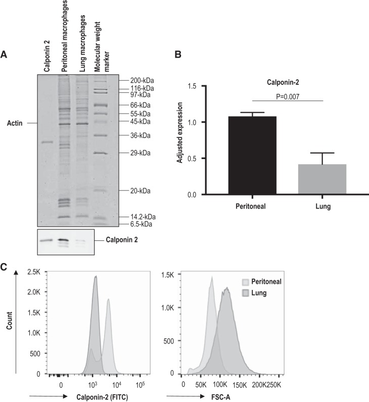
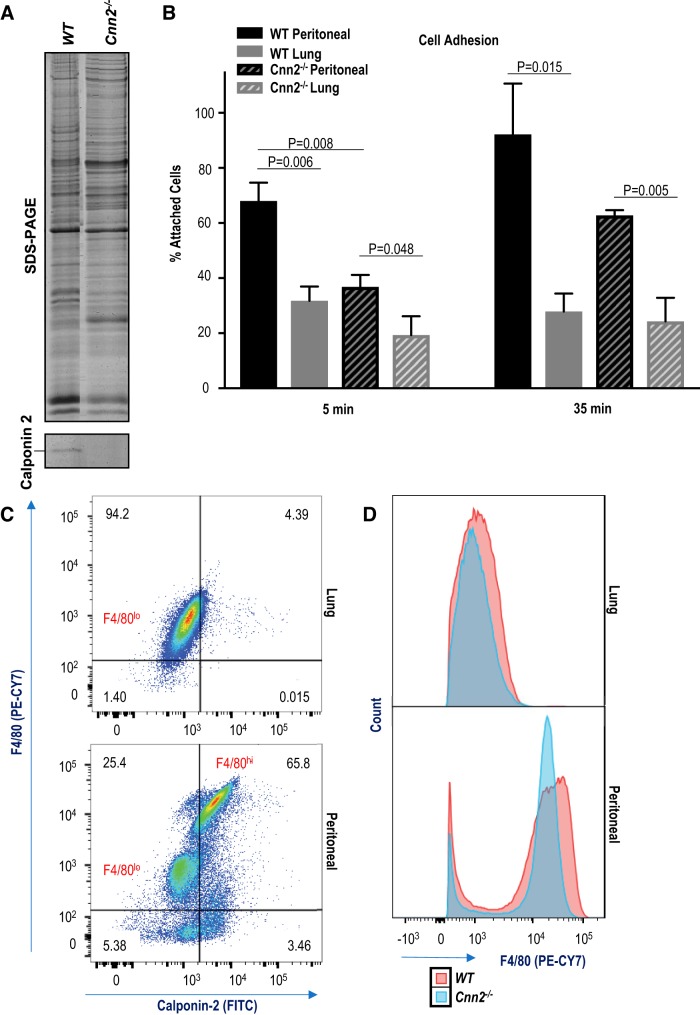
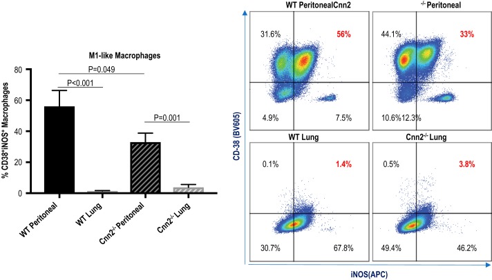
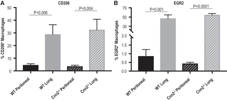
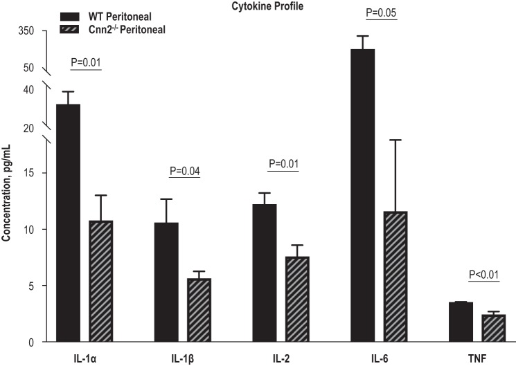
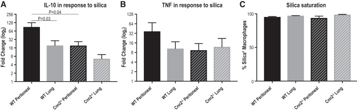
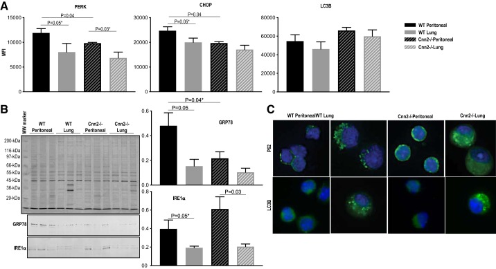
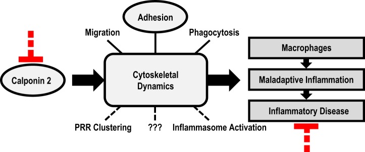
Calponin 2 is an actin cytoskeleton-associated regulatory protein that inhibits the activity of myosin-ATPase and cytoskeleton dynamics. Recent studies have demonstrated that deletion of calponin 2 restricts the proinflammatory activation of macrophages in atherosclerosis and arthritis to attenuate the disease progression in mice. Here we demonstrate that the levels of calponin 2 vary among different macrophage populations, which may reflect their adaptation to specific tissue microenvironment corresponding to specific functional states. Interestingly, lung resident macrophages express significantly lower calponin 2 than peritoneal resident macrophages, which correlates with decreased substrate adhesion and reduced expression of proinflammatory cytokines and a proresolution phenotype. Deletion of calponin 2 in peritoneal macrophages also decreased substrate adhesion and downregulated the expression of proinflammatory cytokines. Providing the first line of defense against microbial invasion while receiving constant exposure to extrinsic antigens, lung macrophages need to maintain a necessary level of activity while limiting exaggerated inflammatory reaction. Therefore, their low level of calponin 2 may reflect an important physiological adaption. Downregulation of calponin 2 in macrophages may be targeted as a cytoskeleton-based novel mechanism, possibly via endoplasmic reticulum stress altering the processing and secretion of cytokines, to regulate immune response and promote quiescence for the treatment of inflammatory diseases.

Keywords: actin; adhesion; cytoskeleton; inflammation; macrophage.


Conflict of interest statement

No conflicts of interest, financial or otherwise, are declared by the authors.

Figures

Fig. 1.
Fig. 1.
Significantly lower level of calponin 2 in lung macrophages than in peritoneal macrophages. A: the levels of calponin 2 in lung and peritoneal macrophages isolated from wild-type mice (n = 4 mice) were determined by immunoblotting. The protein input was normalized by the level of actin. Purified mouse calponin 2 protein was used as a positive control. B: the results were quantified by densitometry analysis using ImageJ software. The level of calponin 2 expression was evaluated relative to the levels of histones that reflect the number of cells. Bars depict means ± SE. C: the expression of calponin 2 in CD45+F4/80+ macrophages was confirmed by flow cytometry using rabbit anti-calponin 2 antibody RAH2 and FITC-conjugated anti-rabbit IgG. The rightward shift in forward scatter area (FSC-A) demonstrates the larger size of freshly isolated lung macrophages when compared with that of peritoneal macrophages.
Fig. 2.
Fig. 2.
Lower levels of calponin 2 lead to reduced substrate adhesion and correlate with decreased expression of F4/80. A: complete deletion of calponin 2 was reconfirmed in spleen harvested from Cnn2−/− mouse with a wild-type (WT) control. B: substrate adhesion of peritoneal and lung macrophages freshly isolated from WT and Cnn2−/− mice (n = 3 mice per group) was determined by direct counting 5 and 35 min after seeding. The data are graphed as the percentage of attached cells out of the total cells expected to attach based on the seeding density and proportion of macrophages of each sample. Bars depict means ± SE. C: the coexpression of F4/80 and calponin 2 in live CD45+F4/80+ macrophages was determined by flow cytometry using anti-F4/80 antibody directly conjugated to PE-CY7 and unlabeled anti-calponin 2 antibody RAH2 in conjunction with FITC-conjugated secondary antibody. The percentage of F4/80+Cnn2+ macrophages is indicated. D: the expression level of F4/80 was compared in WT and Cnn2−/− CD45+F4/80+ macrophages using flow cytometry. Histograms display the overlays of PE-CY7 fluorescent spectra for lung and peritoneal macrophages.
Fig. 3.
Fig. 3.
Lower levels of calponin 2 correlate to reduced proportion of M1-like CD38+iNOS+ macrophages. Flow cytometric analysis was performed on peritoneal and lung macrophages freshly isolated from wild-type (WT) and Cnn2−/− mice (n = 4 mice per group) to determine the expression of M1-like markers CD-38 and inducible nitric oxide synthase (iNOS) in live CD45+F4/80+ cells. The percentages of CD38+iNOS+ macrophages are represented by bar graphs (means ± SE), and the expression pattern of CD38 and iNOS in macrophages is depicted by dot plots.
Fig. 4.
Fig. 4.
Higher expression of M2-like markers CD206 and early growth response protein 2 (EGR2) in lung versus peritoneal macrophages independent of calponin 2. Flow cytometric analysis was performed on peritoneal and lung macrophages freshly isolated from wild-type (WT) and Cnn2−/− mice (n = 3–5 mice per group) to determine the expression of M2-like markers CD-206 and EGR2 in live CD45+F4/80+ cells. A: the percentages of CD206+ macrophages are represented by bar graphs (means ± SE). B: the percentages of EGR2+ macrophages are represented by bar graphs (means ± SE).
Fig. 5.
Fig. 5.
Deletion of calponin 2 results in decreases of IL-1α, IL-1β, IL-6, and TNF in peritoneal macrophages. Total protein was extracted from peritoneal macrophages freshly isolated from wild-type (WT) and Cnn2−/− mice (n = 3 mice per group) and the levels of proinflammatory cytokines IL-1α, IL-1β, IL-6, and TNF were determined using bead-based multiplex immunoassays. Bars depict means ± SE.
Fig. 6.
Fig. 6.
Lower levels of calponin 2 attenuate inflammatory response of macrophages to silica particles. Peritoneal and lung macrophages freshly isolated from wild-type (WT) and Cnn2−/− mice (n = 3 mice per group) were cultured for 72 h with or without 200-nm silica particles (100 μg/mL). A and B: the treated cells were subjected to flow cytometric analysis to examine the expression of IL-10 (A) and TNF (B) in live CD45+F4/80+ macrophages. The fold change in cytokine expression (the ratio of the difference between silica-stimulated and unstimulated cytokine expression over the unstimulated expression) is depicted by bar graphs (means ± SE). C: saturated silica phagocytosis under the experimental condition was confirmed by parallel incubation of the macrophages with green fluorescent silica particles. The percentage of the macrophages that contain silica is depicted by bar graphs (means ± SE).
Fig. 7.
Fig. 7.
Calponin 2 contributes to endoplasmic reticulum (ER) stress response of macrophages. A: flow cytometric analysis was performed on peritoneal and lung macrophages freshly isolated from wild-type (WT) and Cnn2−/− mice (n = 3 mice per group) to determine the expression of ER stress response markers PERK (protein kinase RNA-like endoplasmic reticulum kinase) and CHOP (CCAAT/enhancer-binding protein homologous protein) and the autophagy marker LC3B in live CD45+F4/80+ macrophages. The mean fluorescence intensity (MFI) is represented by bar graphs (means ± SE). *One-tailed Student’s t test. B: the levels of glucose-regulated protein 78 (GRP78) and inositol-requiring enzyme-1α (IRE1α) in lung and peritoneal macrophages isolated from WT and Cnn2−/− mice (n = 3 mice per group) were determined by immunoblotting. The protein input was normalized by the level of actin. C: immunofluorescence microscopic localization of P62 and LC3B (green) was performed on WT and Cnn2−/− lung and peritoneal macrophages. Nuclei were stained with DAPI (blue). Representative images were obtained using a ×40 objective.
Fig. 8.
Fig. 8.
Calponin 2 as a novel target for modulating macrophage function in inflammatory responses. Calponin 2 is a cytoskeleton regulatory protein with a demonstrated function in macrophage migration, adhesion, and phagocytosis and potential roles in pattern recognition receptor (PRR) clustering, inflammasome activation, and other intracellular processes that may regulate the activation and polarization of macrophages implicated in the maladaptive activation in inflammatory diseases. Targeting calponin 2 presents a potentially novel strategy to modulate cytoskeleton-mediated functions and activation of macrophages for the treatment of inflammatory disease.

Similar articles

Cited by

References

    1. Abe M, Takahashi K, Hiwada K. Effect of calponin on actin-activated myosin ATPase activity. J Biochem 108: 835–838, 1990. doi:10.1093/oxfordjournals.jbchem.a123289. - DOI - PubMed
    1. Amit I, Winter DR, Jung S. The role of the local environment and epigenetics in shaping macrophage identity and their effect on tissue homeostasis. Nat Immunol 17: 18–25, 2016. [Erratum in Nat Immunol 18: 246, 2017]. doi:10.1038/ni.3325. - DOI - PubMed
    1. Bain CC, Hawley CA, Garner H, Scott CL, Schridde A, Steers NJ, Mack M, Joshi A, Guilliams M, Mowat AM, Geissmann F, Jenkins SJ. Long-lived self-renewing bone marrow-derived macrophages displace embryo-derived cells to inhabit adult serous cavities. Nat Commun 7: ncomms11852, 2016. doi:10.1038/ncomms11852. - DOI - PMC - PubMed
    1. Bronner DN, Abuaita BH, Chen X, Fitzgerald KA, Nuñez G, He Y, Yin XM, O’Riordan MX. Endoplasmic reticulum stress activates the inflammasome via NLRP3- and caspase-2-driven mitochondrial damage. Immunity 43: 451–462, 2015. doi:10.1016/j.immuni.2015.08.008. - DOI - PMC - PubMed
    1. Burger D, Fickentscher C, de Moerloose P, Brandt KJ. F-actin dampens NLRP3 inflammasome activity via Flightless-I and LRRFIP2. Sci Rep 6: 29834, 2016. doi:10.1038/srep29834. - DOI - PMC - PubMed

Publication types

LinkOut - more resources