Comparative Study
. 2019 Jul 31;14(7):e0220360.
doi: 10.1371/journal.pone.0220360. eCollection 2019.

Comparison of implantation sites for the development of peritoneal metastasis in a colorectal cancer mouse model using non-invasive bioluminescence imaging

Affiliations
Comparative Study

Comparison of implantation sites for the development of peritoneal metastasis in a colorectal cancer mouse model using non-invasive bioluminescence imaging

Abdelkader Taibi et al. PLoS One. .

Abstract

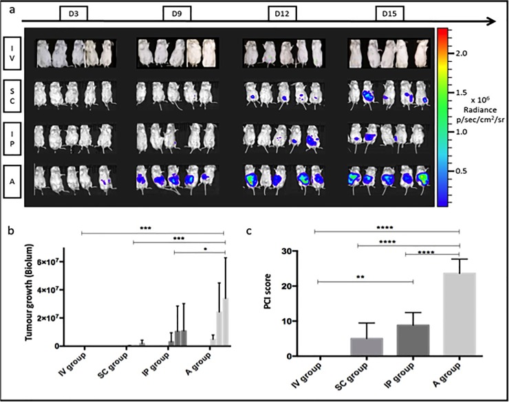
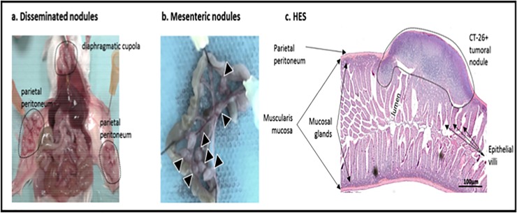
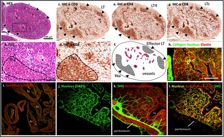
The development of cancer mouse models is still needed for the identification and preclinical validation of novel therapeutic targets in colorectal cancer, which is the third leading cause of cancer-related deaths in Europe. The purpose of this study was to determine the most accurate tumour cell injection method to obtain suitable peritoneal metastasis (PM) for subsequent therapeutic treatments. Here, we grafted murine colon carcinoma CT-26 cells expressing luciferase into immunocompetent BALB-c mice by intravenous injection (IV group), subcutaneous injection (SC group), intraperitoneal injection after peritoneal scratching (A group) or intraperitoneal injection alone (IP group). Tumour growth was monitored by bioluminescence during the first 15 days post-grafting. The peritoneal carcinomatosis index was evaluated macroscopically, histology, immunohistochemistry and multiphoton microscopy were performed in peritoneal tumour tissue. Upon implantation, no tumour growth was observed in the IV group, similar to the non-injected group. Both the IP and SC groups showed intermediate growth rates, but the SC group produced only a single subcutaneous nodule. The A group exhibited the highest tumour growth at 15 days post-surgery. Anatomic and histologic analyses corroborated the existence of various tumour nodules, and multiphoton microscopy was used to evaluate tumour fibrosis-infiltrating cells in a non-pathologic peritoneum. In conclusion, limited PM was obtained by IP injection, whereas IP injection after peritoneal scratching led to an extensive PM murine model for evaluating new therapeutics.


Conflict of interest statement

The authors have declared that no competing interests exist.

Figures

Fig 1
Fig 1. In vivo quantification of tumor growth by bioluminescence tracking of luciferase expressing CT-26 cells over 15 days.
(A) Microphotographs of anesthetized mice for each group inside the bioluminescent imaging box. (B) Tumor growth evaluation based on bioluminescence measurements for the four groups (IV, SC, IP, A) at day 3, day 9, day 12 and day 15 (low values are not always visible on the graph). (C) PCI score at day 15 post-grafting. In summary, the mice having undergone laparotomy surgery (group A: intraperitoneal cell injection after peritoneum aggression) present an extensive tumoral growth with the highest peritoneal carcinomatosis index (PCI). Furthermore, statistically significant differences are observed between the four groups. n = 5 for each group, mean +- SD. * for p value <0.05, ** <0.01, *** <0.001 and ****<0.0001.
Fig 2
Fig 2
Images after mouse laparotomy at day 15 post-grafting, (A) A group showed extensive intraperitoneal tumor growth localized majority on the parietal peritoneum and the diaphragm, and (B) IP group displayed a limited peritoneal metastasis localized on the visceral peritoneum. (C) HES staining in the intestinal bowel with peritoneal tumoral nodule.
Fig 3
Fig 3
(A-B) HES staining and (C-G) immunohistochemistry for CD3, CD4, CD8 of a 15 days grafted CT-26+ nodule in a mouse from IP group. Grafted nodules reveal high number of cells including Tumor infiltrating Lymphocytes (TILs) among a complex tumoral microenvironment (high vascularization, poor fibrosis, dense tissue). Multiphoton imaging of tumoral tissue (H) and normal peritoneum (I-L). v = vessel, LT = T Lymphocyte, LT4 = T4 Lymphocyte, LTc = cytotoxic Lymphocyte, lu = lumen, SHG = second harmonic generation. Scale bar in a applied to c,e,f,g, in b applied to d, and in h applied to i-l.

References

    1. Jayne DG, Fook S, Loi C, Seow-Choen F. Peritoneal carcinomatosis from colorectal cancer. Br J Surg 2002; 89(12):1545–1550. 10.1046/j.1365-2168.2002.02274.x - DOI - PubMed
    1. Russell AH, Tong D, Dawson LE, Wisbeck W. Adenocarcinoma of the proximal colon. Sites of initial dissemination and patterns of recurrence following surgery alone. Cancer 1984; 53(2):360–367. 10.1002/1097-0142(19840115)53:2<360::aid-cncr2820530232>3.0.co;2-u - DOI - PubMed
    1. Russell AH, Tong D, Dawson LE et al. Adenocarcinoma of the retroperitoneal ascending and descending colon: sites of initial dissemination and clinical patterns of recurrence following surgery alone. Int. J. Radiat. Oncol. Biol. Phys. 1983; 9(3):361–365. 10.1016/0360-3016(83)90297-3 - DOI - PubMed
    1. Maggiori L, Elias D. Curative treatment of colorectal peritoneal carcinomatosis: current status and future trends. Eur J Surg Oncol 2010; 36(7):599–603. 10.1016/j.ejso.2010.05.007 - DOI - PubMed
    1. Klaver CEL, Stam R, Sloothaak DAM et al. Colorectal cancer at high risk of peritoneal metastases: long term outcomes of a pilot study on adjuvant laparoscopic HIPEC and future perspectives. Oncotarget 2017; 8(31):51200–51209. 10.18632/oncotarget.17158 - DOI - PMC - PubMed

Publication types