. 2019 Jul 29;94(3):264-278.
doi: 10.1590/abd1806-4841.20199011.

Pemphigus vulgaris

Affiliations

Pemphigus vulgaris

Adriana Maria Porro et al. An Bras Dermatol. .

Abstract

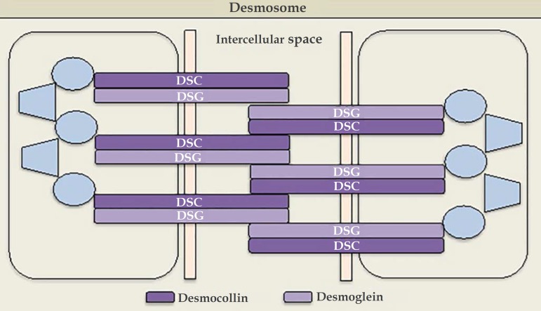
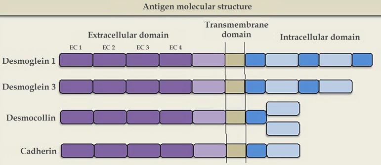
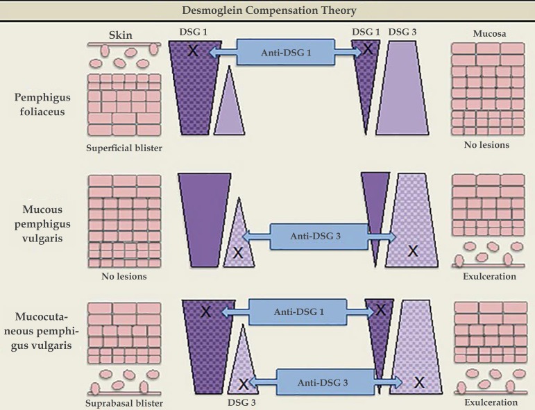
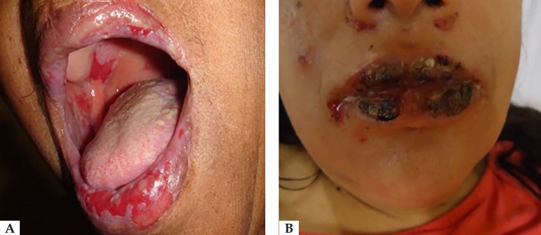
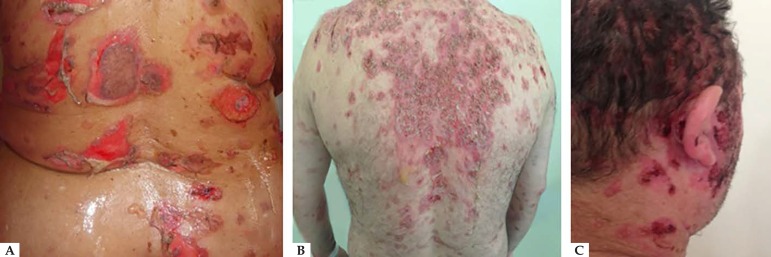
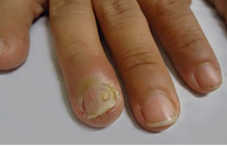
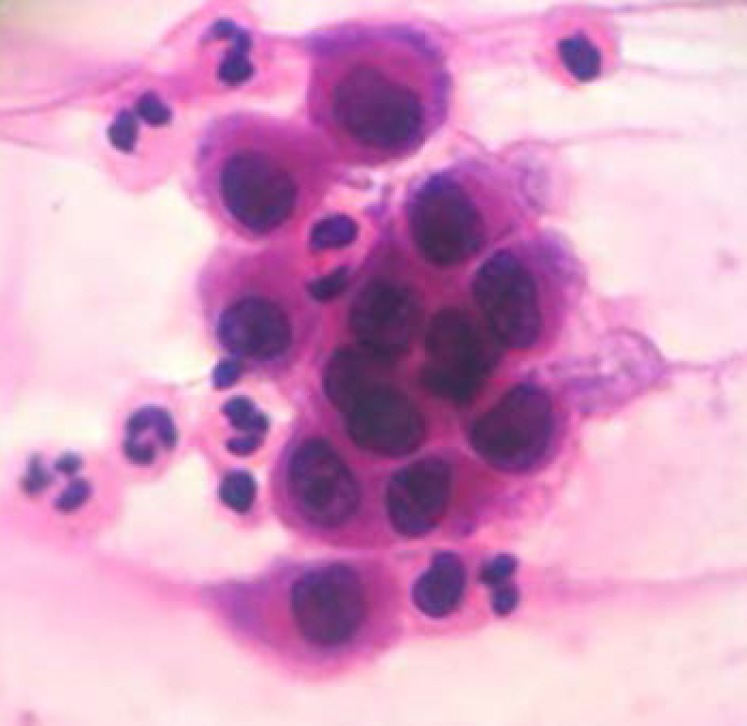
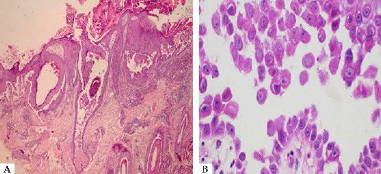
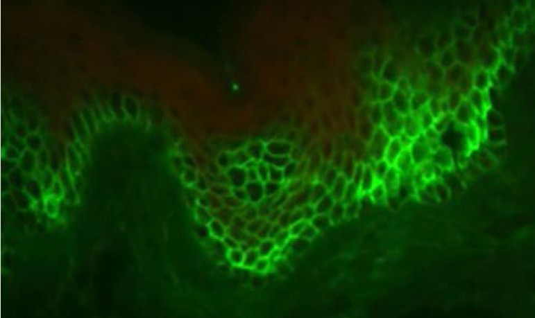
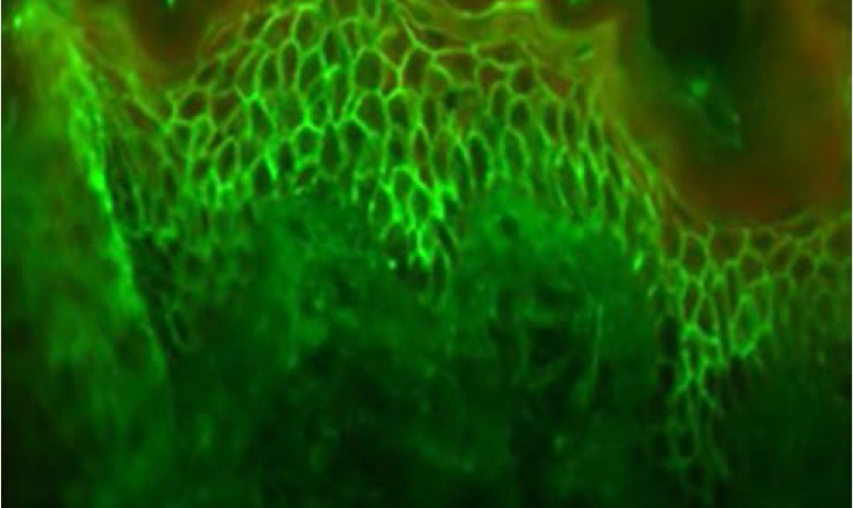
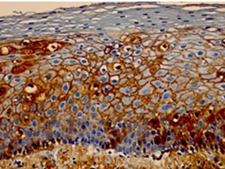
Pemphigus vulgaris is a chronic autoimmune bullous dermatosis that results from the production of autoantibodies against desmogleins 1 and 3. It is the most frequent and most severe form of pemphigus, occurring universally, usually between 40 and 60 years of age. It usually begins with blisters and erosions on the oral mucosa, followed by lesions on other mucous membranes and flaccid blisters on the skin, which can be disseminated. There is a clinical variant, pemphigus vegetans, which is characterized by the presence of vegetating lesions in the large folds of the skin. Clinical suspicion can be confirmed by cytological examination, histopathological examination, and direct and indirect immunofluorescence tests. The treatment is performed with systemic corticosteroids, and immunosuppressive drugs may be associated, among them azathioprine and mycophenolate mofetil. More severe cases may benefit from corticosteroids in the form of intravenous pulse therapy, and recent studies have shown a beneficial effect of rituximab, an anti-CD20 immunobiological drug. It is a chronic disease with mortality around 10%, and septicemia is the main cause of death. Patients need long-term and multidisciplinary follow-up.


Conflict of interest statement

Conflict of Interest: None.

Figures

Figure 1
Figure 1
The desmosomal complex includes desmoglein, desmocollin, and transmembrane and cytoplasmic components
Figure 2
Figure 2
The molecular structure of the pemphigus antigens comprises the extracellular region (EC), with four calcium-dependent cadherin repeats
Figure 3
Figure 3
Desmoglein compensation theory. The different distribution patterns of Dsg1 and Dsg3 in the skin and mucosa are represented. In pemphigus foliaceus, anti-Dsg1 IgG antibodies cause superficial blisters on the skin, as Dsg3 compensates for non-functioning Dsg1 in the deep epidermis; there are no mucosal lesions, since adhesion is mediated mainly by Dsg3. In mucosal pemphigus vulgaris, anti-Dsg3 IgG antibodies do not cause skin damage because Dsg1 compensates for Dsg3 dysfunction; however, there is a mucosal lesion because, unlike the skin, the low concentration of Dsg1 in the mucous membranes is not enough to compensate for Dsg3 dysfunction. In mucocutaneous pemphigus, the presence of anti-Dsg1 and anti-Dsg3 IgG antibodies causes lesions on both the skin and the mucosa
Figure 4
Figure 4
A. Vesicles, blisters, and exulcerations on the lips. Exulcerations in the buccal and palate mucosa; B. Serohematic exulcerations and crusts on the lips
Figure 5
Figure 5
A. Vesicles, blisters, and exulcerations on the dorsum; B. Exulcerations and crusts on the back; C. Exulcerations and crusts on the scalp
Figure 6
Figure 6
PV paronychia
Figure 7
Figure 7
Grouping of acantholytic keratinocytes observed on cytological examination by the Tzanck method (Hematoxylin & eosin, x400)
Figure 8
Figure 8
A - Histopathological examination, showing a blister with suprabasal cleavage level affecting the epidermis and follicular epithelium (Hematoxylin & eosin, x40); B - In detail, acantholytic keratinocytes in the blister content (Hematoxylin & eosin, x400)
Figure 9
Figure 9
Direct immunofluorescence examination showing moderate intensity for the IgG and C3 markers, with intercellular fluorescence distribution, often with predominant location in the lower layers of the epithelium
Figure 10
Figure 10
Indirect immunofluorescence with intercellular intraepithelial IgG
Figure 11
Figure 11
Immunohistochemical examination using an IgG marker (C3 is similar) showing intense intercellular immunoexpression

Similar articles

Cited by

References

    1. Kridin K, Sagi SZ, Bergman R. Mortality and cause of death in Israeli patients with pemphigus. Acta Derm Venereol. 2017;97:607–611. - PubMed
    1. Hsu DY, Brieva J, Sinha AA, Langan SM, Silverberg J. Comorbidities and inpatient mortality for pemphigus in the USA. Br J Dermatol. 2016;174:1290–1298. - PubMed
    1. Di Zenzo G, Amber KT, Sayar BS, Müller EJ, Borradori L. Immune response in pemphigus and beyond: progresses and emerging concepts. Semin Immunopathol. 2016;38:57–74. - PubMed
    1. Mahoney MG, Wang Z, Rothenberger K, Koch PJ, Amagai M, Stanley JR. Explanations for the clinical and microscopic localization of lesions in pemphigus foliaceus and vulgaris. J Clin Invest. 1999;103:461–468. - PMC - PubMed
    1. Pan M, Liu X, Zheng J. The pathogenic role of autoantibodies in pemphigus vulgaris. Clin Exp Dermatol. 2011;36:703–707. - PubMed