

doi: 10.1590/abd1806-4841.20197906.
Scanning electron microscopy of dermatofibroma
Affiliations
- PMID: 31365670
- PMCID: PMC6668944
- DOI: 10.1590/abd1806-4841.20197906
Item in Clipboard
Scanning electron microscopy of dermatofibroma
An Bras Dermatol.
.
Abstract
Dermatofibroma is a proliferation of spindle cells located in the dermis. We used scanning electron microscopy to examine two histologically confirmed lesions and observed preserved collagen bundles in the perilesional area. In the lesional area, the collagen was denser, without formation of bundles. Higher magnification showed collagen with mesh-like appearance similar to stretched tufts of cotton. Very high magnification evidenced the tufts of cotton and spindle cells measuring 2 to 12 microns.
Conflict of interest statement
Conflict of interest: None.
Figures
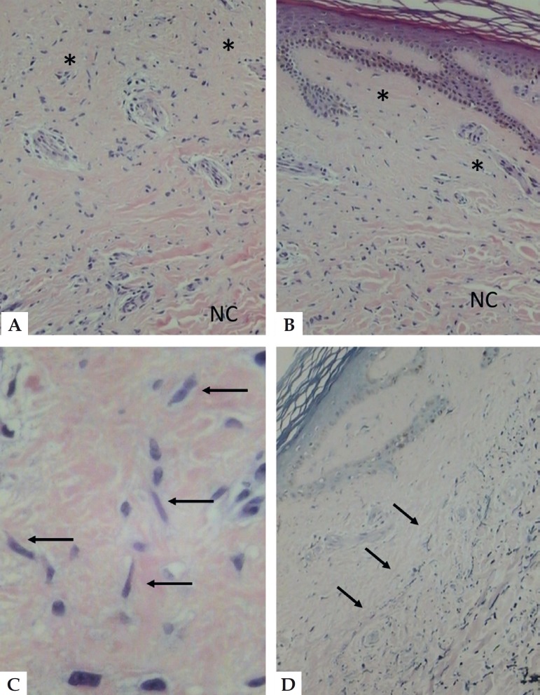
Light Microscopy - A - Collagen homogenization with cell
proliferation (asterisks) with normal collagen in the lower portion (NC)
(hematoxylin & eosin, x150). B - similar aspect in the
lesion border, with involvement of the papillary dermis (hematoxylin
& eosin, x150). C - detail of the fusiform cells
(arrows) (hematoxylin & eosin, x400). D - diminished
elastic fibers in the homogenous area, with clear demarcation of normal
elastic tissue (arrows) in the lower portion (Weigert’s stain, x150)
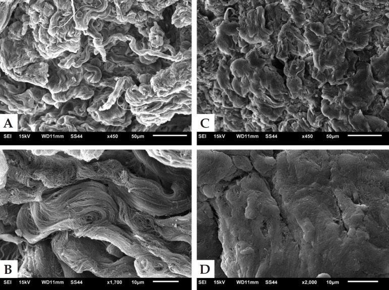
Scanning electron microscopy. A - Low magnification of the
perilesional area with normal collagen bundles (x450). B -
High er magnification of normal collagen bundles (x1.700).
C - Low mag nification of the lesional area with
compact tufted collagen (x450). D - Higher magnification of
affected compacted collagen (x2.000)
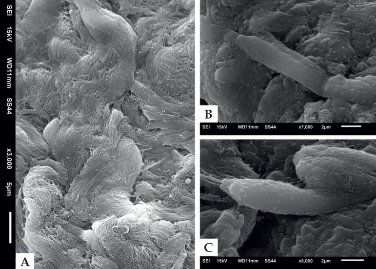
Scanning electron microscopy. A - Detail of the affected
area with tufted collagen without bundle formation (x3.000). B e
C - Detail of fusiform cells, measuring 2 to 12 microns
(x7,000 and x8,000)
References
-
- Barker SM, Winkelmann RK. Inflammatory lymphadenoid reactions with dermatofibroma/histiocytoma. J Cutan Pathol. 1986;13:222–226. - PubMed
-
- Song Y, Sakamoto F, Ito M. Characterization of factor XIIIa+ dendritic cells in dermatofibroma: Immunohistochemical, electron and immunoelectron microscopical observations. J Dermatol Sci. 2005;39:89–96. - PubMed
-
- Gonzales S, Duarte I. Benign fibrous histiocytoma of the skin. A morphologic study of 290 cases. Pathol Res Pract. 1982;174:379–391. - PubMed
MeSH terms
LinkOut - more resources
Full Text Sources
Medical
