. 2019 Jul 30;11(8):692.
doi: 10.3390/v11080692.

BST2/Tetherin Overexpression Modulates Morbillivirus Glycoprotein Production to Inhibit Cell-Cell Fusion

Affiliations

BST2/Tetherin Overexpression Modulates Morbillivirus Glycoprotein Production to Inhibit Cell-Cell Fusion

James T Kelly et al. Viruses. .

Abstract

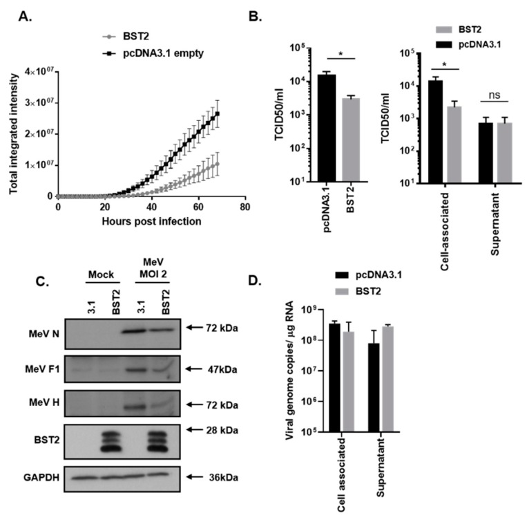
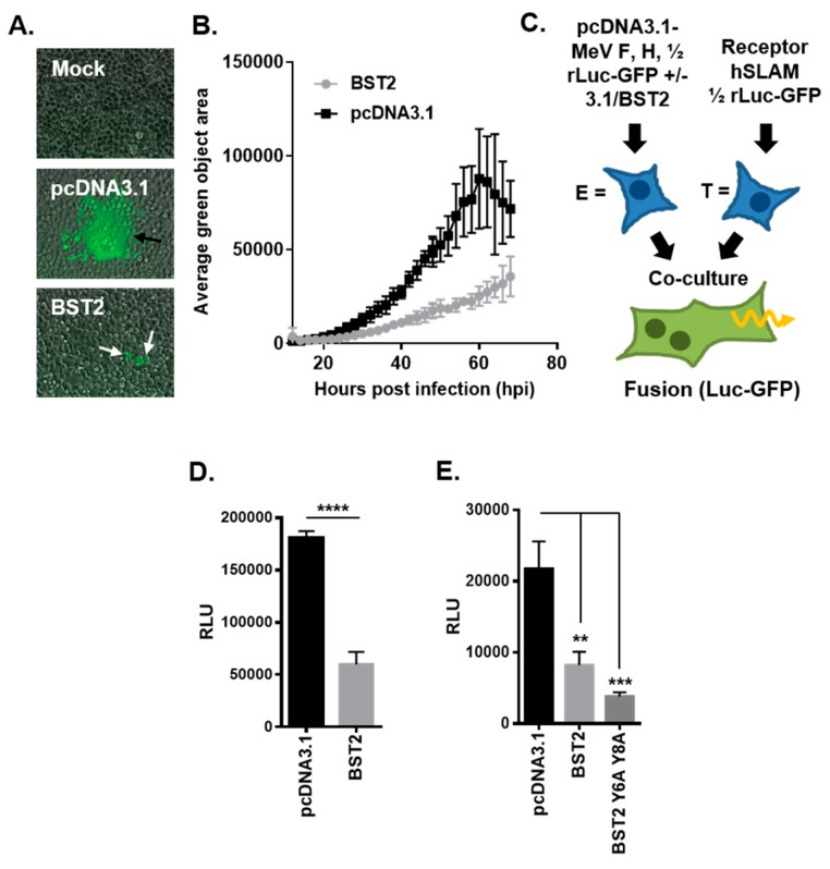
The measles virus (MeV), a member of the genus Morbillivirus, is an established pathogen of humans. A key feature of morbilliviruses is their ability to spread by virus-cell and cell-cell fusion. The latter process, which leads to syncytia formation in vitro and in vivo, is driven by the viral fusion (F) and haemagglutinin (H) glycoproteins. In this study, we demonstrate that MeV glycoproteins are sensitive to inhibition by bone marrow stromal antigen 2 (BST2/Tetherin/CD317) proteins. BST2 overexpression causes a large reduction in MeV syncytia expansion. Using quantitative cell-cell fusion assays, immunolabeling, and biochemistry we further demonstrate that ectopically expressed BST2 directly inhibits MeV cell-cell fusion. This restriction is mediated by the targeting of the MeV H glycoprotein, but not other MeV proteins. Using truncation mutants, we further establish that the C-terminal glycosyl-phosphatidylinositol (GPI) anchor of BST2 is required for the restriction of MeV replication in vitro and cell-cell fusion. By extending our study to the ruminant morbillivirus peste des petits ruminants virus (PPRV) and its natural host, sheep, we also confirm this is a broad and cross-species specific phenotype.

Keywords: BST2; MeV; Morbillivirus; PPRV; haemagluttinin; measles; tetherin.


Conflict of interest statement

The authors declare no conflict of interest, financial or otherwise.

Figures

Figure 1
Figure 1
Measles virus (MeV) replication is sensitive to bone marrow stromal antigen 2 (BST2). 293-hSLAMs, transfected with the indicated pcDNA3.1-based expression construct for 24 h, were infected with MeV-GFP at a multiplicity of infection (MOI) of 2 (A–D) and virus replication assayed periodically using an Incucyte imager for 72 h with GFP as a marker (A). In equivalent experiments mean, virus titres were calculated at 48 h post infection by TCID50 analysis of triplicate biological samples of supernatant and cell-associated virus (B); (left panel, sum of total virus titres and right panel, individual fractions). At equivalent time points infected cells were lysed in RIPA buffer and analyzed using western blot to quantify viral protein production (C) or, separately, the RNA was extracted, and the genome was quantified using strand-specific qPCR (D). Error bars indicate standard deviation of the mean. Statistical significance is as follows: *, p < 0.05.
Figure 2
Figure 2
MeV cell–cell fusion is inhibited by BST2. (A/B) The average green object size (syncytia) in high MOI (2), MeV-GFP infected 293-hSLAM cells, transfected with the indicated pcDNA3.1-based expression constructs, was quantified at the indicated times post infection using an Incucyte imager. (A) Representative micrographs taken at 24 h post infection (black arrows, typical syncytia in pcDNA3.1 transfected; white arrows, smaller foci with pcDNA3.1-BST2). (C) The MeV cell–cell fusion assay involves separate transfection/treatment of effector E and target T populations prior to co-culturing, as indicated. Fused cells can be visualized and quantified due to reconstitution of the rLuc-GFP reporter. (D) The HEK293T MeV effector cells, expressing MeV fusion (F) and haemagglutinin (H), were cotransfected with the indicated pcDNA3.1-based expression constructs before cell fusion was quantified (by assaying Renilla luciferase activity 16 h after co-culturing with hSLAMF1-positive target cells). (E) The MeV F/H effector cells were cotransfected with pcDNA3.1, BST2 wt, or BST2 Y6,8A expression constructs and cell–cell fusion assayed, as described previously. Error bars indicate standard deviation (B) or error (D/E) of the mean. Statistical significance is as follows: **, p < 0.005; ***, p < 0.0005; ****, p < 0.0001.
Figure 3
Figure 3
The MeV haemagglutinin glycoprotein is targeted by BST2. (A) Vero-hSLAM cells, cotransfected with the indicated pcDNA3.1-based expression constructs, and analyzed by immunofluorescence microscopy. Representative micrographs are shown together with merged images to demonstrate colocalization of labeled proteins. (B/C) 293-hSLAMs were transfected with 500 ng of pcDNA3.1-MeV nucleocapsid (N) or H and either pcDNA3.1, or, increasing amounts of pcDNA3.1-BST2 (ranging from 0 to 1.6 μg) and analyzed by western blot. The total mass of transfected DNA remained constant.
Figure 4
Figure 4
The BST2 GPI anchor is required for restriction of MeV replication. (A) The human BST2 protein is a membrane anchored protein composed of various domains (TM, transmembrane; GPI, glycosyl-phosphatidylinositol). (B/D) 293-hSLAMs transfected with pcDNA3.1, BST2, ∆-GPI, or ∆-TM were cotransfected with either 500 ng of pcDNA3.1 (B) or 250 ng each of pcDNA3.1-MeV F and H (D) and analyzed by western blot. The total mass of transfected DNA remained constant. (C) The HEK293T MeV effector cells were cotransfected with the indicated pcDNA3.1-based expression constructs and cell fusion quantified by assaying Renilla luciferase activity 16 h after co-culturing with hSLAMF1-positive target cells. (E) The Vero-hSLAM cells, cotransfected with the indicated pcDNA3.1-based expression constructs, and analyzed by immunofluorescence microscopy. Representative micrographs are shown together with merged images to demonstrate colocalization of labeled proteins. (F) 293-hSLAMs, transfected with the indicated pcDNA3.1-based expression construct for 24 h, were infected with MeV-GFP and total virus replication assayed at 72 h. Error bars indicate standard deviation (F) or error (C) of the mean. Statistical significance is as follows: *, p < 0.05; **, p < 0.005; ****, p < 0.0001.
Figure 5
Figure 5
Morbilliviruses are broadly targeted by mammalian BST2 proteins. (A) Phylogenetic analysis of the amino acid sequences of a range of mammalian BST2 proteins including those examined in this study (highlighted in red). (B/E) Vero-hSLAM cells, cotransfected with the indicated pcDNA3.1-based expression constructs, and analyzed by immunofluorescence microscopy. Representative micrographs are shown together with merged images to demonstrate colocalization of labeled proteins. (C/F) HEK293T cells transfected with pcDNA3.1-, H.s BST2, O.a BST2A, or O.a BST2B, were cotransfected with either 500 ng of pcDNA3.1 (C) or 250 ng each of pcDNA3.1 peste des petits ruminants virus (PPRV) F and H (F) and analyzed by western blot. The total mass of transfected DNA remained constant. (D/G) HEK293T PPRV (D) or MeV (G) effector cells were cotransfected with the indicated pcDNA3.1-based expression constructs and cell fusion quantified by assaying Renilla luciferase activity 16 h after co-culturing with ovine or human SLAMF1-positive target cells, respectively. Error bars indicate standard error of the mean. Statistical significance is as follows: ***, p < 0.0005; ****, p < 0.0001.
Figure 6
Figure 6
H protein production and trafficking is inhibited in PPRV-infected BST2-overexpressing cells. PPRV H distribution is modified in BST2-transfected infected cells. (A) Vero cSLAM cells were transfected with plasmids expressing the indicated BST2 protein (or mock transfected) before being infected with PPRV Tu00 at an MOI of 0.1. Cells were fixed at 16 h post infection before staining with relevant antibodies (PPRV H, red and BST2, green). Micrographs were taken on a Leica confocal microscope with line of interest analysis of colocalization performed using the inbuilt software. The white boxed insets in the merged panels reflect the region chosen for analysis in the “zoom” panel (right), with the x-axis on the colocalization graphs representing the 15 µm white lines indicated on the adjacent zoomed images. (B) A zoomed image of a single large BST2-expressing syncytia indicating the approximate position of the plasma membrane.

Similar articles

Cited by

References

    1. World Health Orgnization Measles (Fact Sheet No 286) [(accessed on 24 July 2019)]; Available online: www.who.int/mediacentre/factsheets/fs286/en/
    1. El Najjar F., Schmitt A.P., Dutch R.E. Paramyxovirus glycoprotein incorporation, assembly and budding: A three way dance for infectious particle production. Viruses. 2014;6:3019–3054. doi: 10.3390/v6083019. - DOI - PMC - PubMed
    1. Muhlebach M.D., Mateo M., Sinn P.L., Prufer S., Uhlig K.M., Leonard V.H., Navaratnarajah C.K., Frenzke M., Wong X.X., Sawatsky B., et al. Adherens junction protein nectin-4 is the epithelial receptor for measles virus. Nature. 2011;480:530–533. doi: 10.1038/nature10639. - DOI - PMC - PubMed
    1. Ludlow M., McQuaid S., Milner D., de Swart R.L., Duprex W.P. Pathological consequences of systemic measles virus infection. J. Pathol. 2015;235:253–265. doi: 10.1002/path.4457. - DOI - PubMed
    1. White R.G., Boyd J.F. The effect of measles on the thymus and other lymphoid tissues. Clin. Exp. Immunol. 1973;13:343–357. - PMC - PubMed

Publication types

MeSH terms

LinkOut - more resources