. 2019 Jul 30;12(15):2429.
doi: 10.3390/ma12152429.

Dental Implants with Anti-Biofilm Properties: A Pilot Study for Developing a New Sericin-Based Coating

Affiliations

Dental Implants with Anti-Biofilm Properties: A Pilot Study for Developing a New Sericin-Based Coating

Paolo Ghensi et al. Materials (Basel). .

Abstract

Aim: several strategies have been tested in recent years to prevent bacterial colonization of dental implants. Sericin, one of the two main silk proteins, possesses relevant biological activities and also literature reports about its potential antibacterial properties, but results are discordant and not yet definitive. The aim of this study was to evaluate the effectiveness of different experimental protocols in order to obtain a sericin-based coating on medical grade titanium (Ti) able to reduce microbial adhesion to the dental implant surface.

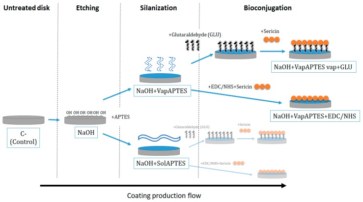
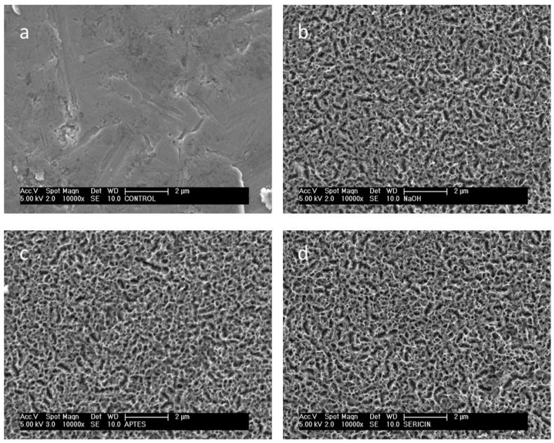
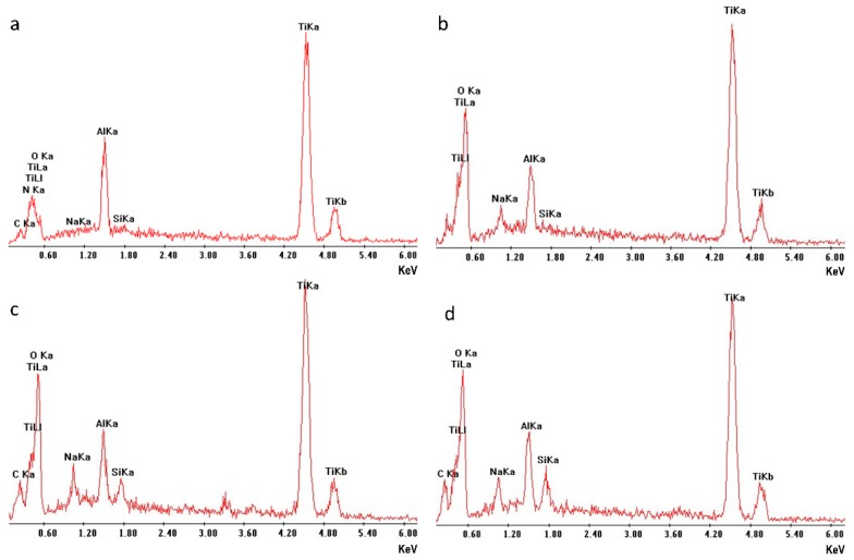
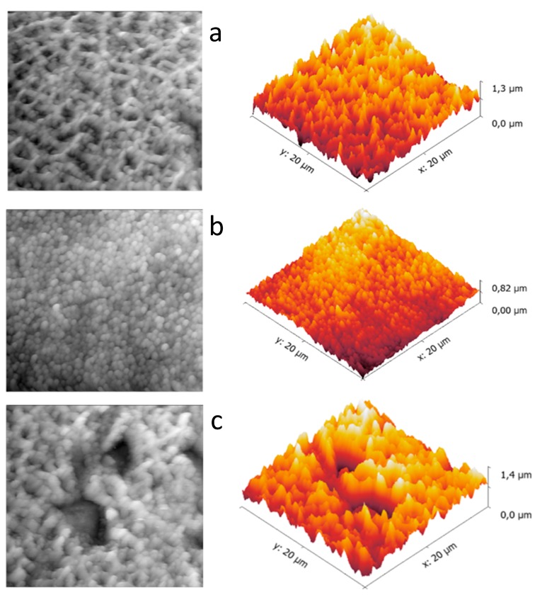
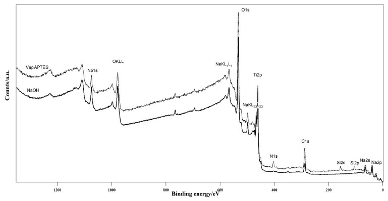
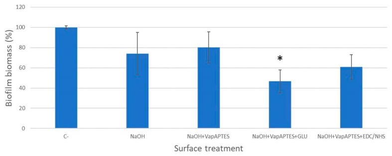
Materials and methods: different strategies for covalent bonding of sericin to Ti were pursued throughout a multi-step procedure on Ti-6Al-4V disks. The surface of grade 5 Ti was initially immersed in NaOH solution to obtain the exposure of functional -OH groups. Two different silanization strategies were then tested using aminopropyltriethoxysilane (APTES). Eventually, the bonding between silanized Ti-6Al-4V and sericin was obtained with two different crosslinking processes: glutaraldehyde (GLU) or carbodiimide/N-Hydroxy-succinimide (EDC/NHS). Micro-morphological and compositional analyses were performed on the samples at each intermediate step to assess the most effective coating strategy able to optimize the silanization and bioconjugation processes. Microbiological tests on the coated Ti-6Al-4V disks were conducted in vitro using a standard biofilm producer strain of Staphylococcus aureus (ATCC 6538) to quantify the inhibition of microbial biofilm formation (anti-biofilm efficacy) at 24 hours.

Results: both silanization techniques resulted in a significant increase of silicon (Si) on the Ti-6Al-4V surfaces etched with NaOH. Differences were found between GLU and EDC/NHS bioconjugation strategies in terms of composition, surface micro-morphology and anti-biofilm efficacy. Ti-6Al-4V samples coated with GLU-bound sericin after silanization obtained via vapor phase deposition proved that this technique is the most convenient and effective coating strategy, resulting in a bacterial inhibition of about 53% in respect to the uncoated Ti-6Al-4V disks.

Conclusions: The coating with glutaraldehyde-bound sericin after silanization in the vapor phase showed promising bacterial inhibition values with a significant reduction of S. aureus biofilm. Further studies including higher number of replicates and more peri-implant-relevant microorganisms are needed to evaluate the applicability of this experimental protocol to dental implants.

Keywords: Ti-6Al-4V; bioconjugation; biofilm; coating; dental implants; titanium (Ti).


Conflict of interest statement

The authors declare no conflict of interest. The funders had no role in the design of the study; in the collection, analyses, or interpretation of data; in the writing of the manuscript, or in the decision to publish the results.

Figures

Figure 1
Figure 1
Diagram of the four investigated coating strategies obtained by combining different silanization (vapor-phase deposition, VapAPTES or organic/aqueous solvent, SolAPTES) and bio conjugations (glutaraldehyde (GLU) or carbodiimide/N-Hydroxy-succinimide (EDC/NHS)) process after a preliminary etching phase (NaOH).
Figure 2
Figure 2
Micromorphology of the disk surface at different production step: (a) untreated control, (b) after etching (NaOH), (c) after etching and silanization (NaOH + VapAPTES), (d) after etching, silanization and sericin bioconjugation with glutaraldehyde (NaOH + VapAPTES + GLU). Images (c,d) are representative also for the micromorphology of disks subjected to the alternative silanization (SolAPTES) and bioconjugation (EDC/NHS) processes respectively. Scanning electron microscopy, secondary electrons detector signal, original magnification 10,000×.
Figure 3
Figure 3
Surface compositional analysis of Ti-6Al-4V disks subjected to different surface treatments: (a) untreated control, (b) after etching (NaOH), (c) after etching and silanization by vapour deposition (NaOH + VapAPTES), (d) after etching and silanization by solvent deposition (NaOH + SolAPTES). Sodium (Na) and oxygen (O) are introduced by the etching phase and Silicon (Si) by silanization phase. Energy dispersive spectroscopy. Primary electron beam energy was set to 15 KeV. Spectra are collected over an area of 25 µm2. Integration time 200 s.
Figure 4
Figure 4
Nano-morphology (2D images on the left and 3D reconstructions on the right) of the disk surface at different production step: (a) after etching and silanization (NaOH + VapAPTES), (b) after etching, silanization and sericin bioconjugation with glutaraldehyde (NaOH + VapAPTES + GLU), (c) after etching, silanization and sericin bioconjugation with EDC/NHS (NaOH + VapAPTES + NDC/NHS). Atomic force microscopy, semi-contact mode.
Figure 5
Figure 5
Comparison of the survey spectra of the samples after NaOH etching but before functionalization (NaOH) and after aminopropyltriethoxysilane (APTES) (VapAPTES) functionalization. Successful attachment of APTES is evidenced by the presence of nitrogen and silicon species, which were not present on the bare Ti-6Al-4V surface.
Figure 6
Figure 6
Biofilm biomass (%) of Staphylococcus aureus ATCC 6538 formed at 24 h of incubation on the surface of Ti-6Al-4V disks subjected to different surface treatments. Data are normalized to untreated controls (C−). *p < 0.05.

Similar articles

Cited by

References

    1. Lang N.P., Berglundh T., on Behalf of Working Group 4 of the Seventh European Workshop on Periodontology Periimplant diseases: Where are we now?—Consensus of the seventh european workshop on periodontology. J. Clin. Periodontol. 2011;38:178–181. doi: 10.1111/j.1600-051X.2010.01674.x. - DOI - PubMed
    1. Do T., Devine D., Marsh P.D. Oral Biofilms: Molecular Analysis, Challenges, and Future Prospects in Dental Diagnostics. Clin. Cosmet. Investig. Dent. 2013;5:11–19. doi: 10.2147/ccide.s31005. - DOI - PMC - PubMed
    1. Derks J., Tomasi C. Peri-Implant Health and Disease. A Systematic Review of Current Epidemiology. J. Clin. Periodontol. 2015;42:S158–S171. doi: 10.1111/jcpe.12334. - DOI - PubMed
    1. Quirynen M., De Soete M., Van Steenberghe D. Infectious Risks for Oral Implants: A Review of the Literature. Clin. Oral Implants Res. 2002;13:1–19. doi: 10.1034/j.1600-0501.2002.130101.x. - DOI - PubMed
    1. Quirynen M., Vogels R., Peeters W., Van Steenberghe D., Naert I., Haffajee A. Dynamics of Initial Subgingival Colonization of “Pristine” Peri-Implant Pockets. Clin. Oral Implants Res. 2006;17:25–37. doi: 10.1111/j.1600-0501.2005.01194.x. - DOI - PubMed

LinkOut - more resources