. 2019 Jul 30;8(8):256.
doi: 10.3390/antiox8080256.

Protective Effects of Dendropanax morbifera against Cisplatin-Induced Nephrotoxicity without Altering Chemotherapeutic Efficacy

Affiliations

Protective Effects of Dendropanax morbifera against Cisplatin-Induced Nephrotoxicity without Altering Chemotherapeutic Efficacy

Ji Su Kim et al. Antioxidants (Basel). .

Abstract

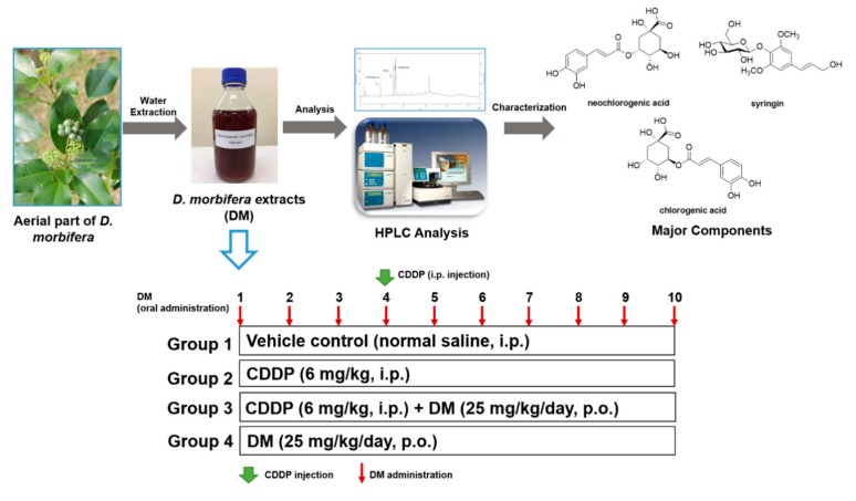
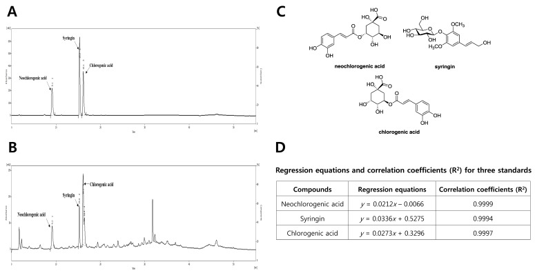
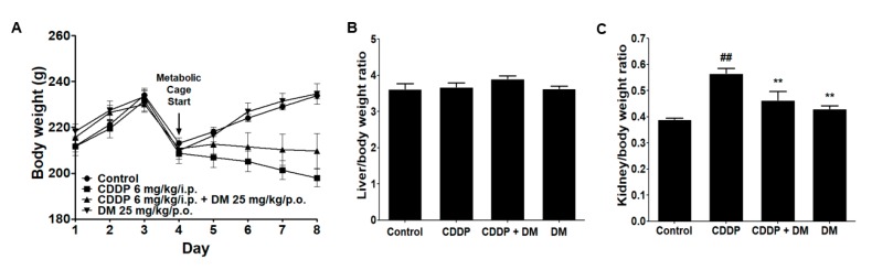
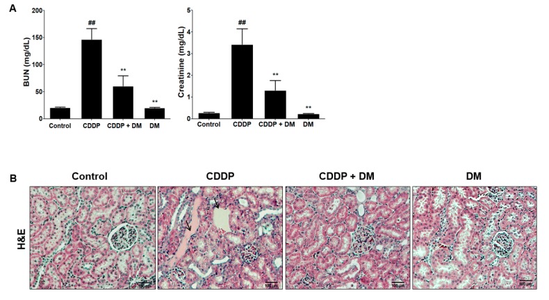
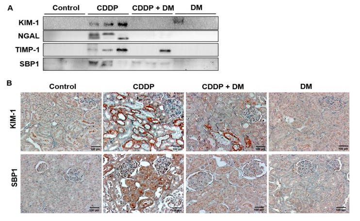
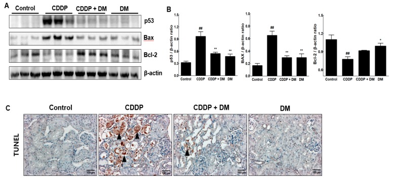
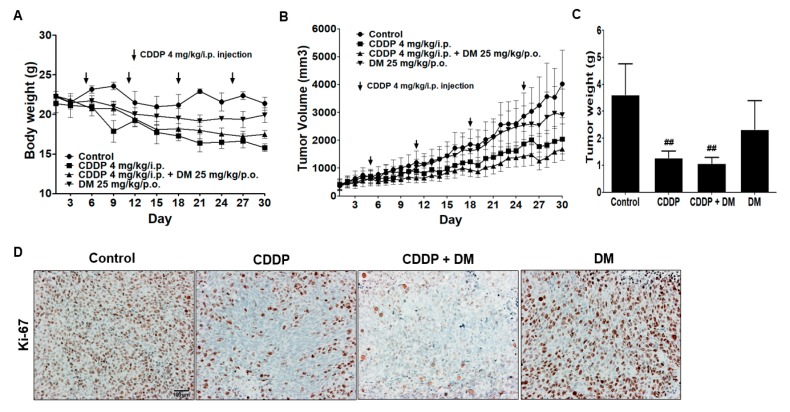
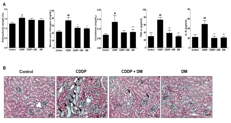
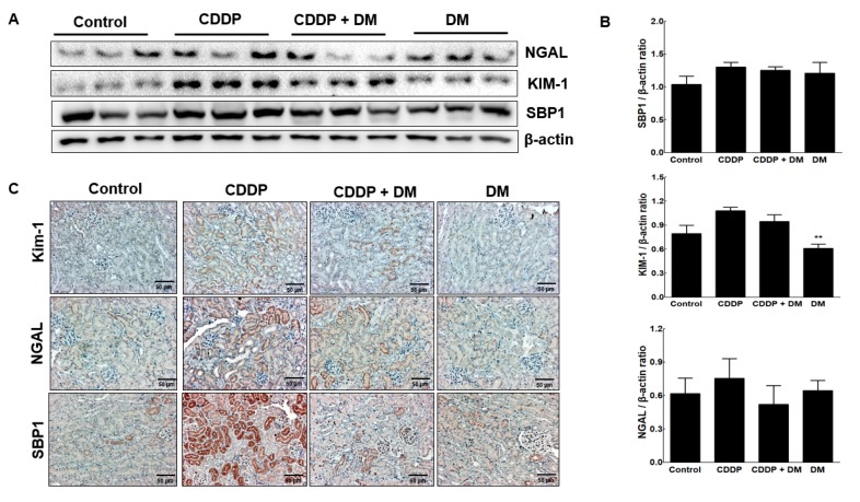
Use of the chemotherapeutic agent cisplatin (CDDP) in cancer patients is limited by the occurrence of acute kidney injury (AKI); however, no protective therapy is available. We aimed to investigate the renoprotective effects of Dendropanax morbifera water extract (DM) on CDDP-induced AKI. Male Sprague-Dawley rats (six animals/group) received: Vehicle (control); CDDP (6 mg/kg, intraperitoneally (i.p.); DM (25 mg/kg, oral); or DM + CDDP injection. CDDP treatment significantly increased blood urea nitrogen (BUN), serum creatinine (sCr), and pro-inflammatory cytokines (IL-6 and TNF-α), and severely damaged the kidney architecture. Urinary excretion of protein-based AKI biomarkers also increased in the CDDP-treated group. In contrast, DM ameliorated CDDP-induced AKI biomarkers. It markedly protected against CDDP-induced oxidative stress by increasing the activity of endogenous antioxidants and reducing the levels of pro-inflammatory cytokines (IL-6 and TNF-α). The protective effect of DM in the proximal tubules was evident upon histopathological examination. In a tumor xenograft model, administration of DM enhanced the chemotherapeutic activity of CDDP and exhibited renoprotective effects against CDDP-induced nephrotoxicity without altering chemotherapeutic efficacy. Our data demonstrate that DM may be an adjuvant therapy with CDDP in solid tumor patients to preserve renal function.

Keywords: Dendropanax morbifera; antioxidants; chemotherapy; cisplatin; renoprotective effect; xenograft model.


Conflict of interest statement

The authors declare no conflict of interest.

Figures

Figure 1
Figure 1
Preparation of Dendropanax morbifera water extract (DM) and experimental design of cisplatin (CDDP)-induced nephrotoxicity in rats. Rats were randomly divided into four groups: The control group, which received normal saline; the CDDP (6 mg/kg, single intraperitoneal (i.p.) injection) group; the CDDP (6 mg/kg, single i.p. injection) + DM (25 mg/kg/day) group; and the DM (25 mg/kg/day) group. The DM was administered orally for 10 days.
Figure 2
Figure 2
Phytochemical characterization of DM by HPLC analysis. (A) HPLC-UV chromatogram for DM; (B) HPLC-UV chromatogram for three standard compounds (neochlorogenic acid, syringin, and chlorogenic acid); (C) chemical structures of three major compounds in DM. (D) Regression equations and correlation coefficients (R2) for the three standard compounds. HPLC analysis was carried out via gradient elution on a Phenomenex Kinetex C18 column (150 × 4.6 mm, 5 µm). The flow rate, column oven temperature, and UV wavelength for detection were set at 1 mL/min, 30 °C, and 254 nm, respectively.
Figure 3
Figure 3
Effects of DM on changes in body, liver, and kidney weights in rats treated with CDDP. (A) Body weight changes in CDDP-treated rats. (B) Liver weight in CDDP-treated rats. (C) Kidney weight in CDDP-treated rats. All values represent the mean ± Standard Deviation (SD) of six rats per group. Statistical analyses were performed by one-way ANOVA followed by Tukey’s HSD (honest significant difference) post hoc test for multiple comparisons. ## p < 0.01 compared to the control group; ** p < 0.01 compared to the CDDP-treated group.
Figure 4
Figure 4
Effects of DM on CDDP-induced nephrotoxicity in rats. (A) Serum biochemical parameters were measured in CDDP-treated rats. Values are the mean ± SD of six rats per group. Statistical analyses were performed using one-way ANOVA followed by Tukey’s HSD post hoc test for multiple comparisons. ## p < 0.01 compared to the control group; ** p < 0.01 compared to the CDDP-treated group. (B) H&E staining of representative kidney tissues from all experimental groups. Black arrows indicate renal tissue necrosis and infiltration in the proximal tubules. Magnification ×100.
Figure 5
Figure 5
Effect of DM on acute kidney injury biomarkers in CDDP-treated rats. (A) Changes in the urinary excretion of KIM-1, NGAL, TIMP-1, and SBP1. The Western blot results represent three separate experiments. (B) Immunohistochemical staining of KIM-1 and SBP1 in the kidney of the rat. Magnification ×100.
Figure 6
Figure 6
Effect of DM on antioxidant enzyme activities and pro-inflammatory cytokine levels in CDDP-treated rats. The activities of superoxide dismutase (SOD) and catalase were measured in the kidney of rats. Pro-inflammatory cytokines levels were measured in the serum of rats. Values are the mean ± SD of six rats per group. Statistical analyses were performed using one-way ANOVA followed by Tukey’s HSD post hoc test for multiple comparisons. # p < 0.05 and ## p < 0.01 compared to the control group; * p < 0.05 and ** p < 0.01 compared to the CDDP-treated group.
Figure 7
Figure 7
Effects of DM on apoptosis in the kidney of CDDP-treated rats. (A) The expression of p53, Bax, and Bcl-2 in the kidney of CDDP-treated rats was measured by Western blot analysis. β-Actin expression was used as the loading control. The Western blot results represent three separate experiments. Each group of blots is the same exposure of a gel; each experiment was repeated more than three times. (B) Representative graphs indicated the fold changes of Western blot data. Values are the mean ± SD of triplicate experiments. Statistical analyses were performed using one-way ANOVA followed by Tukey’s HSD post hoc test for multiple comparisons. ## p < 0.01 compared to the control group; * p < 0.05 and ** p < 0.01 compared to the CDDP-treated group. (C) Detection of apoptosis via TUNEL assay in the kidney tissues of rats. Black arrowheads represent a high expression of TUNEL-positive cells. Magnification ×100.
Figure 8
Figure 8
Effect of DM on CDDP-induced nephrotoxicity in tumor xenograft mice. (A) Effect of DM on body weight changes in tumor xenograft mice treated with CDDP. (B) Each graph indicates the mean tumor volumes in tumor xenograft mice after treatment with drugs for 30 days. (C) Each bar represents the mean tumor weight. Statistical analyses were performed using one-way ANOVA followed by Tukey’s HSD post hoc test for multiple comparisons. ## p < 0.01 compared to the control group. (D) Immunohistochemical staining for Ki-67 in tumor tissues. Magnification ×100, Scale bar = 100 μm. Representative immunohistochemical images were captured under a 40× objective lens.
Figure 9
Figure 9
Protective effect of DM on CDDP-induced nephrotoxicity in tumor xenograft mice transfected with HCT-116 cells. (A) Kidney weight, nephrotoxicity biomarkers, and inflammatory cytokine levels were measured in the serum of tumor-bearing mice treated with CDDP. Values are the mean ± SD of six mice per group. Statistical analyses were performed using one-way ANOVA followed by Tukey’s HSD post hoc test for multiple comparisons. ## p < 0.05 and ## p < 0.01 compared to the control group; * p < 0.05 and ** p < 0.01 compared to the CDDP-treated group. (B) Representative histology of H&E-stained kidney sections in the experimental groups of a tumor xenograft mouse model. At 31 days, CDDP-induced acute kidney injury (AKI) mice presented an enlarged cortex with glomerular sclerosis (arrowheads). The cortex from CDDP-treated mice exhibited a normal-sized renal cortex and a lower incidence of tubular and medulla injury and displayed a normal histological structure of thin tubules and collecting ducts. Images are representative of three animals per experimental group (magnification ×100, bar = 100 μm).
Figure 10
Figure 10
Protective effect of DM on nephrotoxicity biomarkers in CDDP-induced nephrotoxicity in tumor xenograft mice transfected with HCT-116 cells. (A) Nephrotoxicity biomarkers were measured in the kidney of mice. Representative Western blots of NGAL, KIM-1, and SBP1 from triplicate experiments are shown. (B) Densitometric analysis showed the expression of SBP1, KIM-1, and NGAL in the kidney of mice with CDDP-induced nephrotoxicity. ** p < 0.01 compared to the CDDP-treated group. (C) Representative immunohistochemical staining of KIM-1, NGAL, and SBP1 in the kidney of mice with CDDP-induced nephrotoxicity. Original magnification ×100, scale bar: 50 μm.

References

    1. Siddik Z.H. Cisplatin: Mode of cytotoxic action and molecular basis of resistance. Oncogene. 2003;22:7265–7279. doi: 10.1038/sj.onc.1206933. - DOI - PubMed
    1. Wang D., Lippard S.J. Cellular processing of platinum anticancer drugs. Nat. Rev. Drug Discov. 2005;4:307–320. doi: 10.1038/nrd1691. - DOI - PubMed
    1. Cepeda V., Fuertes M.A., Castilla J., Alonso C., Quevedo C., Pe’rez J.M. Biochemical mechanisms of cisplatin cytotoxicity. Anticancer Agents Med. Chem. 2007;7:3–18. doi: 10.2174/187152007779314044. - DOI - PubMed
    1. Meyer K.B., Madias N.E. Cisplatin nephrotoxicity. Miner. Electrolyte Metab. 1994;20:201–213. - PubMed
    1. Miller R.P., Tadagavadi R.K., Ramesh G., Reeves W.B. Mechanisms of Cisplatin Nephrotoxicity. Toxins. 2010;2:2490–2518. doi: 10.3390/toxins2112490. - DOI - PMC - PubMed

LinkOut - more resources