. 2019 Jul 31;11(503):eaaw3768.
doi: 10.1126/scitranslmed.aaw3768.

Therapeutically relevant engraftment of a CRISPR-Cas9-edited HSC-enriched population with HbF reactivation in nonhuman primates

Affiliations

Therapeutically relevant engraftment of a CRISPR-Cas9-edited HSC-enriched population with HbF reactivation in nonhuman primates

Olivier Humbert et al. Sci Transl Med. .

Abstract

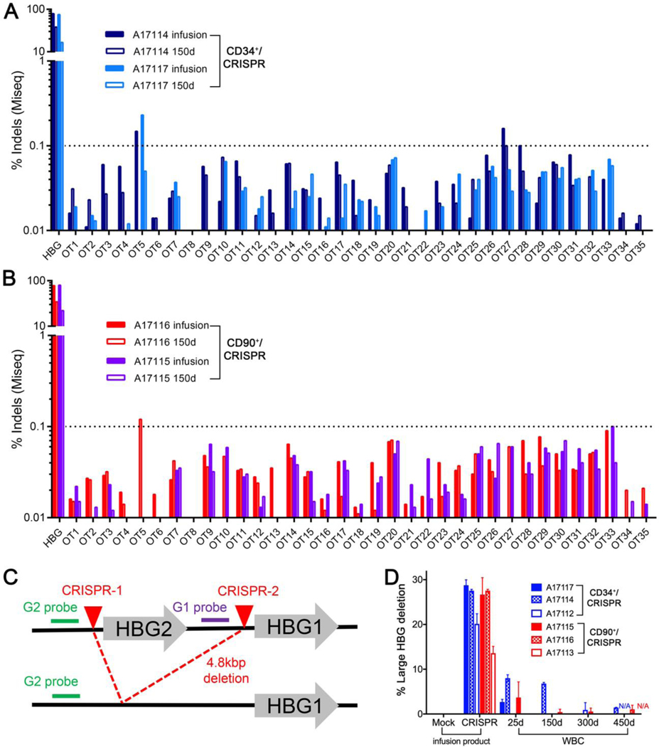
Reactivation of fetal hemoglobin (HbF) is being pursued as a treatment strategy for hemoglobinopathies. Here, we evaluated the therapeutic potential of hematopoietic stem and progenitor cells (HSPCs) edited with the CRISPR-Cas9 nuclease platform to recapitulate naturally occurring mutations identified in individuals who express increased amounts of HbF, a condition known as hereditary persistence of HbF. CRISPR-Cas9 treatment and transplantation of HSPCs purified on the basis of surface expression of the CD34 receptor in a nonhuman primate (NHP) autologous transplantation model resulted in up to 30% engraftment of gene-edited cells for >1 year. Edited cells effectively and stably reactivated HbF, as evidenced by up to 18% HbF-expressing erythrocytes in peripheral blood. Similar results were obtained by editing highly enriched stem cells, defined by the markers CD34+CD90+CD45RA-, allowing for a 10-fold reduction in the number of transplanted target cells, thus considerably reducing the need for editing reagents. The frequency of engrafted, gene-edited cells persisting in vivo using this approach may be sufficient to ameliorate the phenotype for a number of genetic diseases.


Figures

Fig. 1.
Fig. 1.. Recapitulation of HPFH genotype in NHP HSPCs.
(A) Schematic of β-globin locus with CCAAT repressor motifs (underlined), putative BCL11a binding sequence TGACCA (green box), and CRISPR/Cas9 target site (red arrow). Sequences highlighted in orange show HPFH sites for 13-nt deletion [−114/−102] and −117 G/A substitution. (B) Deletion profile in NHP CD34+ edited cells (animal A17117) at 4 days post-editing. Colored boxes show identified distinct deletions relative to the total sequencing pool, and the white portion shows all combined deletions contributing less than 1%. The 13-nt deletion is on top in the dark blue box. (C) Genomic sequences of the most common deletions from (B) with length of deletions on the left in nucleotides (nt). Boxes highlight 8-nt microhomology repeats. (D) Immunophenotypic separation of HSPC subsets after CD34+ enrichment (A17117). (E) Flow cytometric validation of the indicated sorted HSPCs subsets from (D). (F) HBG editing efficiency measured at 24 hours post-treatment in sorted subsets from (E). (G) Contribution of 13-nt HPFH deletion relative to all other deletions in edited subsets from (C). (H) CFC assay of CD34+ and HSPC subsets taken at 24 hours after mock electroporation (left) or CRISPR/Cas9 RNP (right) treatment (n=1). CFU=Colony-forming unit, CFU-M=macrophages, CFU-G=granulocytes, CFU-GM=granulocyte/macrophage, BFU-E=erythroid. In panels F and G, results are means and standard deviations from A17117 and A17114. *denotes statistical significance (two-tailed unpaired t-test, P<0.05) of the difference in 13-nt deletion in CD90+ subset as compared to CD34+ cells.
Fig. 2.
Fig. 2.. Tracking of HBG editing in all transplanted animals.
(A) Editing efficiency was measured in PB white blood cells from transplanted animals. Inset shows magnification for the early time points. (B) Editing efficiency measured in the infusion product of transplanted animals at 3–5 days after treatment (n=3). (C) Flow cytometric validation of CD90+ and CD90-sorted populations after CD34+ enrichment (A17116). (D) CFC assay of sorted populations from (C) before editing (left) and 24 hours after editing (right). CFU-M=macrophages, CFU-G=granulocytes, CFU-GM=granulocyte/macrophage, BFU-E=erythroid. (E) Normalized frequency of the 13-nt HPFH deletion in reactions from (B), n=3. * denotes statistical significance (two-tailed unpaired t-test, P<0.05) of the difference in 13-nt deletion in CD90+ subset as compared to CD34+. Deletion profile of CD34+/CRISPR animal A17114 (F) or CD90+/CRISPR animal A17116 (G) after transplant. In panels F and G, colored boxes show identified distinct deletions relative to the total sequencing pool, and the white portion shows all combined deletions contributing less than 1%. The 13-nt deletion is on top in the dark blue box. (H) Contribution of the 13-nt HPFH deletion in the same animals as in (A) after normalization of all deletion frequencies to 100%.
Fig. 3.
Fig. 3.. HbF response in transplanted animals.
(A) Longitudinal measurement of F-cell frequency in PB of transplanted animals from CD34+ (blue) and CD90+ (red) cohorts as compared to historical transplant controls (gray) and one untransplanted control (black). (B) Longitudinal HPLC measurement of γ-globin protein expression (calculated as γ/(γ+β) globin) in the same animals as in panel A. Globin expression was calculated from the area of each eluted peak in HPLC chromatograms. (C) Linear correlation analysis of F-cell frequency (y-axis, red) or γ-globin protein expression (y-axis, purple) and HBG editing frequency (x-axis, determined by TIDE) at 200 to 300 days post-transplant.
Fig. 4.
Fig. 4.. BM analysis from treated animals at 6 months post-transplant.
(A) Immunophenotypic characterization of CD34+ subsets from two CD34+ animals (middle) and two CD90+ animals (right) as compared to untransplanted animal controls (left). (B) Representative CFC assays obtained from BM of transplanted animals from panel A. CFU-M=macrophages, CFU-G=granulocytes, CFU-GM=granulocyte/macrophage, BFU-E=erythroid. (C) HBG editing in BM-sorted populations from two CD34+ animals (left) and two CD90+ animals (right). (D) Deletion profile in different HSPC subsets of BM from the same animals as in (C). N/A=not available. (E) HBG editing measured in different cell lineages of BM from the same animals as in (C). (F) Deletion profile in different cell lineages of BM from the same animals as in (C). In panels D, F, all deletion frequencies were normalized to 100%; colored boxes show distinct deletions, and the white portion shows all combined deletions contributing less than 1%. The 13-nt deletion is on top in the dark blue box.
Fig. 5.
Fig. 5.. Safety of HBG-directed CRISPR/Cas9 gene editing.
(A-B) Frequencies of predicted off-target (OT) sites determined by next generation sequencing analysis of infusion products (CD34+ or CD90+ cells) and of PB sampled at about 150 days post-transplant in CD34+/CRISPR animals (A17117/A17114) (A) and in CD90+/CRISPR animals (A17115/A17116) (B). (C) Schematic representation of CRISPR/Cas9-induced 4.8 kbp deletion with position of primers and probes. (D) Droplet digital PCR (ddPCR) quantification of the 4.8 kbp deletion in the infusion product (CD34+ or CD90+ cells, left) and from PB of the indicated animals at different time points post-transplant. WBC = white blood cells. Results show means and standard deviations from 2 or 3 technical replicates.

References

    1. Modell B, Darlison M, Global epidemiology of haemoglobin disorders and derived service indicators. Bull World Health Organ 86, 480–487 (2008). - PMC - PubMed
    1. Leonard A, Tisdale JF, Stem cell transplantation in sickle cell disease: therapeutic potential and challenges faced. Expert Rev Hematol 11, 547–565 (2018). - PMC - PubMed
    1. Esrick EB, Bauer DE, Genetic therapies for sickle cell disease. Semin Hematol 55, 76–86 (2018). - PubMed
    1. Forget BG, Molecular basis of hereditary persistence of fetal hemoglobin. Ann N Y Acad Sci 850, 38–44 (1998). - PubMed
    1. Gilman JG, Mishima N, Wen XJ, Stoming TA, Lobel J, Huisman TH, Distal CCAAT box deletion in the A gamma globin gene of two black adolescents with elevated fetal A gamma globin. Nucleic Acids Res 16, 10635–10642 (1988). - PMC - PubMed

Publication types