. 2019 Jul 31;10(1):3427.
doi: 10.1038/s41467-019-11363-x.

A one-gate elevator mechanism for the human neutral amino acid transporter ASCT2

Affiliations

A one-gate elevator mechanism for the human neutral amino acid transporter ASCT2

Alisa A Garaeva et al. Nat Commun. .

Abstract

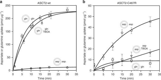
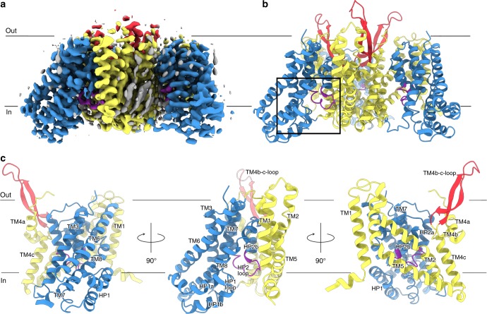
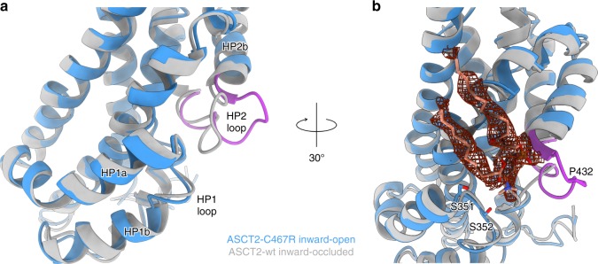
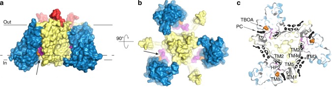
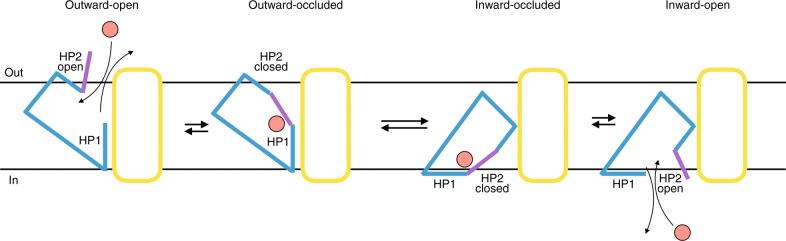
The human Alanine Serine Cysteine Transporter 2 (ASCT2) is a neutral amino acid exchanger that belongs to the solute carrier family 1 (SLC1A). SLC1A structures have revealed an elevator-type mechanism, in which the substrate is translocated across the cell membrane by a large displacement of the transport domain, whereas a small movement of hairpin 2 (HP2) gates the extracellular access to the substrate-binding site. However, it has remained unclear how substrate binding and release is gated on the cytoplasmic side. Here, we present an inward-open structure of the human ASCT2, revealing a hitherto elusive SLC1A conformation. Strikingly, the same structural element (HP2) serves as a gate in the inward-facing as in the outward-facing state. The structures reveal that SLC1A transporters work as one-gate elevators. Unassigned densities near the gate and surrounding the scaffold domain, may represent potential allosteric binding sites, which could guide the design of lipidic-inhibitors for anticancer therapy.


Conflict of interest statement

The authors declare no competing interests.

Figures

Fig. 1
Fig. 1
Transport activity of reconstituted purified ASCT2wt (a) and ASCT2C467R (b). Curves show the counterflow of external radioactively-labelled and internal unlabelled l-glutamine (squares, n = 6 for ASCT2wt, n = 3 for ASCT2C467R); counterflow of l-glutamine in presence of 0.6 mM DL-TBOA (upward triangle, n = 4); counterflow of l-aspartate (circles, n = 3 for ASCT2wt, n = 5 for ASCT2C467R); and counterflow of l-aspartate in presence of 0.5 mM DL-TBOA (downward triangles, n = 3). Data points and error bars represent means and s.e.m. from n biologically independent experiments. Small schemes depict proteoliposomes with internal and external compound compositions used in respective experiments. Source data are provided as a Source Data file
Fig. 2
Fig. 2
Inward-open structure of ASCT2C467R. Cryo-EM map of ASCT2C467R obtained in presence of TBOA at 3.6 Å resolution (a) and respective model shown as ribbon (b). c Protomer of ASCT2 with labelled structural elements is shown as ribbon, rotations are indicated. The scaffold domains are coloured in yellow, the extracellular antenna in red, the transport domains in blue, the HP2 loop in purple, and non-protein densities in grey (see also Supplementary Fig. 7). The membrane boundary is indicated and the location of the substrate binding site as shown in Fig. 3 is highlighted by a black rectangle in panel b
Fig. 3
Fig. 3
Substrate-binding site and movement of the HP2 loop. a, b Superposition of the inward-open ASCT2C467R in presence of TBOA (shown in blue with the HP2 loop in purple) and the substrate-bound inward-occluded ASCT2wt (shown in grey, PDB-ID: 6GCT), demonstrating the movement of HP2. b Non-protein density (shown as red mesh at 3σ) located near HP1 and HP2 with a modelled diundecylphosphidylcholine and putatively coordinating residues shown as sticks. The lipid head group is positioned between the HP1 and HP2 loops and its negative charge appears to be stabilised by the helix dipole of hairpin helix HP2b. a, b Structures were superimposed on the transport domain
Fig. 4
Fig. 4
Accessibility of the substrate-binding site and lipid densities. a, b Surface representation of the ASCT2C467R structure in presence of TBOA, with the transport domain in blue, the scaffold in yellow, the antennae in red, the HP2 loop in purple and TBOA in orange. The open HP2 gate provides access to the binding site from the cytoplasm (indicated by arrows). c Slice through the surface representation of ASCT2C467R (level is indicated by dashed line in panel a) with putative lipid densities surrounding the scaffold domain shown in black and TBOA as orange balls (see also Supplementary Fig. 7)
Fig. 5
Fig. 5
One-gate elevator transport mechanism. Schematic representation of the transport cycle of SLC1A transporters, with the scaffold domain in yellow, the transport domain in blue, the extra- and intracellular gate (HP2 loop) in purple and the transported amino acid in pink

References

    1. Slotboom DJ, Konings WN, Lolkema JS. Structural features of the glutamate transporter family. Microbiol. Mol. Biol. Rev. 1999;63:293–307. - PMC - PubMed
    1. Garaeva AA, et al. Cryo-EM structure of the human neutral amino acid transporter ASCT2. Nat. Struct. Mol. Biol. 2018;25:515–521. doi: 10.1038/s41594-018-0076-y. - DOI - PubMed
    1. Kanner BI, Sharon I. Active transport of L-glutamate by membrane vesicles isolated from rat brain. Biochemistry. 1978;17:3949–3953. doi: 10.1021/bi00612a011. - DOI - PubMed
    1. Kanai Y, et al. The SLC1 high-affinity glutamate and neutral amino acid transporter family. Mol. Aspects Med. 2013;34:108–120. doi: 10.1016/j.mam.2013.01.001. - DOI - PubMed
    1. Wise DR, Thompson CB. Glutamine addiction: a new therapeutic target in cancer. Trends Biochem. Sci. 2010;35:427–433. doi: 10.1016/j.tibs.2010.05.003. - DOI - PMC - PubMed

Publication types

MeSH terms

Substances