Comparison of percentage free PSA, MRI and GaPSMA PET scan for diagnosing cancer prostate in men with PSA between 4 and 20 ng/ml
- PMID: 31367071
- PMCID: PMC6639993
- DOI: 10.4103/iju.IJU_91_19
Comparison of percentage free PSA, MRI and GaPSMA PET scan for diagnosing cancer prostate in men with PSA between 4 and 20 ng/ml
Abstract
Introduction: We compared the diagnostic accuracy of percentage free prostate-specific antigen (PSA), multiparametric magnetic resonance imaging (mpMRI), and gallium-68 prostate-specific membrane antigen positron emission tomography (Ga-PSMA PET) to detect cancer prostate in men with PSA between 4 and 20 ng/ml in prebiopsy settings.
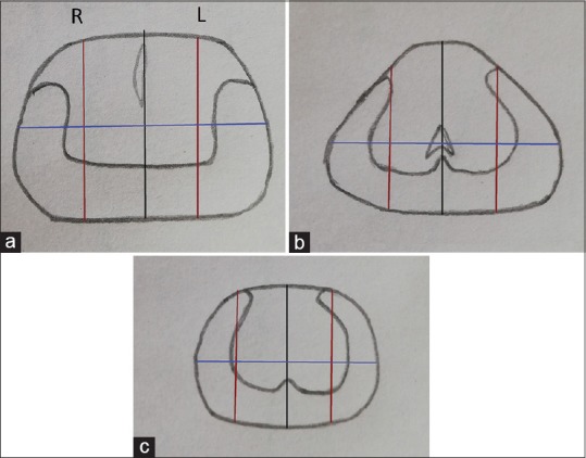
Materials and methods: This prospective study evaluated men with PSA values between 4 and 20 ng/ml, and all patients underwent percentage free PSA estimation, mpMRI, and Ga-PSMA PET scan, followed by cognitive fusion/registration biopsy along with systematic 12-core biopsy to detect cancer prostate. The diagnostic accuracy of percentage free PSA, mpMRI, and Ga-PSMA PET scan was compared with results of cognitive fusion/registration biopsy.
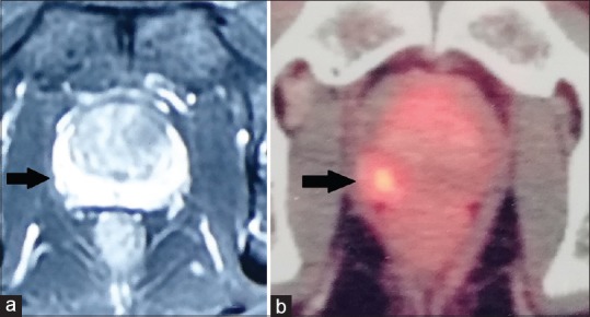
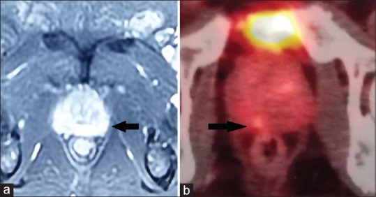
Results: A total of 15 patients were included, of which 11 had an identifiable lesion on imaging and 9 had malignancy on the final histopathology report. The sensitivity, specificity, positive predictive value, negative predictive value (NPV), and diagnostic accuracy of mpMRI were 62.5%, 71.4%, 71.4%, 62.5%, and 66.6%, respectively, and that of Ga-PSMA PET scan were 88.8%, 66.6%, 80%, 80%, and 80%, respectively. The sensitivity of detection of clinically significant cancers for Ga-PSMA was higher (100%) compared to MRI (33.3%). However, Ga-PSMA also detected a greater number of insignificant lesions as compared to MRI.
Conclusion: Ga-PSMA PET scan has high NPV and accuracy in predicting presence of cancer and can also be used to direct specific biopsy cores during systematic biopsy.
Conflict of interest statement
Conflicts of interest: There are no conflicts of interest.
Figures
References
-
- Roehl KA, Antenor JA, Catalona WJ. Serial biopsy results in prostate cancer screening study. J Urol. 2002;167:2435–9. - PubMed
-
- Djavan B, Ravery V, Zlotta A, Dobronski P, Dobrovits M, Fakhari M, et al. Prospective evaluation of prostate cancer detected on biopsies 1, 2, 3 and 4: When should we stop? J Urol. 2001;166:1679–83. - PubMed
-
- Gretzer MB, Partin AW. PSA levels and the probability of prostate cancer on biopsy. Eur Urol Suppl. 2002;1:21–7.
-
- Hamoen EHJ, de Rooij M, Witjes JA, Barentsz JO, Rovers MM. Use of the prostate imaging reporting and data system (PI-RADS) for prostate cancer detection with multiparametric magnetic resonance imaging: A diagnostic meta-analysis. Eur Urol. 2015;67:1112–21. - PubMed
-
- Schoots IG, Roobol MJ, Nieboer D, Bangma CH, Steyerberg EW, Hunink MG. Magnetic resonance imaging-targeted biopsy may enhance the diagnostic accuracy of significant prostate cancer detection compared to standard transrectal ultrasound-guided biopsy: A systematic review and meta-analysis. Eur Urol. 2015;68:438–50. - PubMed
LinkOut - more resources
Full Text Sources
Research Materials
Miscellaneous