MRI - ultrasound fusion guided biopsy of the prostate: lesion volume as a predictor of cancer in patients with repeat biopsies
- PMID: 31367072
- PMCID: PMC6640000
- DOI: 10.4103/iju.IJU_49_19
MRI - ultrasound fusion guided biopsy of the prostate: lesion volume as a predictor of cancer in patients with repeat biopsies
Abstract
Introduction: The objective was to analyze the diagnostic value of multiparametric magnetic resonance imaging (MRI) prostate lesion volume (PLV) and its correlation with the subsequent MRI-ultrasound (MRI-US) fusion biopsy results.
Materials and methods: Between March 2014 and July 2016, 150 men underwent MRI-US fusion biopsies at our institution. All suspicious prostate lesions were graded according to the Prostate Imaging Reporting and Data System (PIRADS) and their volumes were measured. These lesions were subsequently biopsied. All data were prospectively collected and retrospectively analyzed. The PLV of all suspicious lesions was correlated with the presence of cancer on the final MRI-US fusion biopsy. The sensitivity, specificity, positive predictive value (PPV), and negative predictive value (NPV) were calculated.
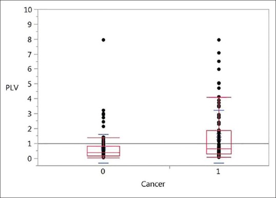
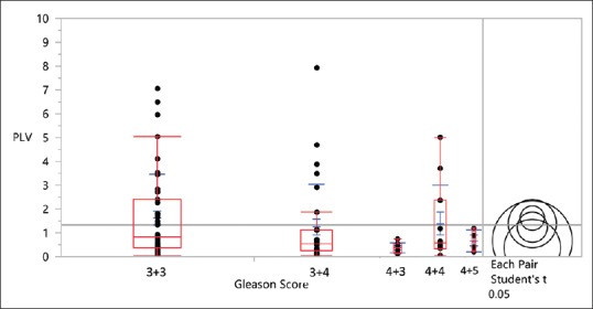
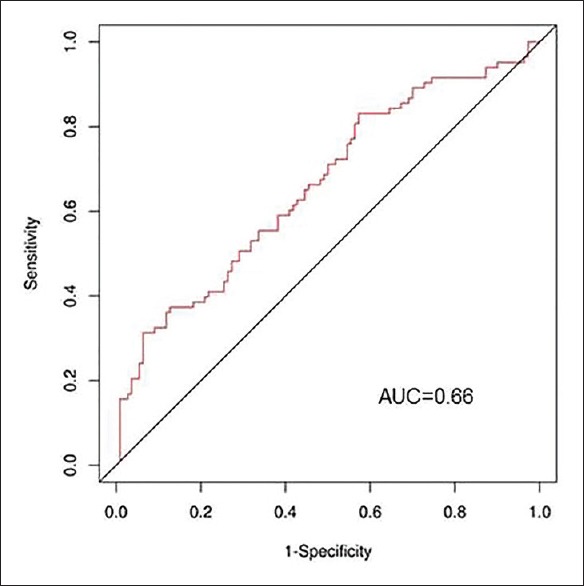
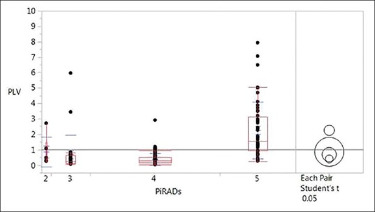
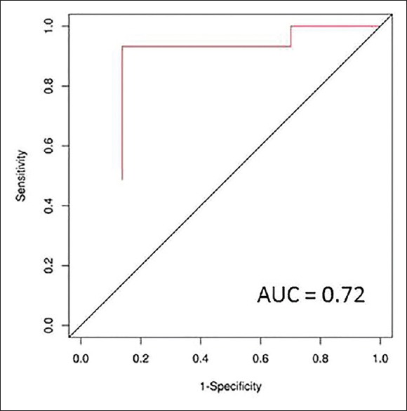
Results: There were 206 suspicious lesions identified in 150 men. The overall cancer detection rate was 102/206 (49.5%). The mean PLV for benign lesions was 0.63 ± 0.94 cm3 versus 1.44 ± 1.76 cm3 for cancerous lesions (P < 0.01). There was a statistically significant difference between the PLV of PIRADS 5 lesions when compared to PIRADS 4, 3, and 2 lesions (P < 0.0001, < 0.0001, and 0.006, respectively). The area under the curve for volume in predicting prostate cancer (PCa) was 0.66. The optimal volume for predicting PCa was 0.26 cm3 with a sensitivity, specificity, PPV, and NPV of 80.7%, 42.7%, 41.2%, and 74.6%, respectively.
Conclusion: PLV may serve as a useful measure to triage patients prior to MRI-US fusion biopsy and help better understand the limits of this technology for individual patients.
Conflict of interest statement
Conflicts of interest: There are no conflicts of interest.
Figures
References
-
- American Cancer Society. Vancouver reference style. Key Statistics for Prostate Cancer. [Last updated on 2016 Mar 11; Last accessed on 2016 Sep 14]. Available from: http://www.cancer.org/cancer/prostatecancer/detailedguide/prostate-cance... .
-
- Levine MA, Ittman M, Melamed J, Lepor H. Two consecutive sets of transrectal ultrasound guided sextant biopsies of the prostate for the detection of prostate cancer. J Urol. 1998;159:471–5. - PubMed
-
- Chen ME, Troncoso P, Johnston DA, Tang K, Babaian RJ. Optimization of prostate biopsy strategy using computer based analysis. J Urol. 1997;158:2168–75. - PubMed
-
- Roehrborn CG, Pickens GJ, Sanders JS. Diagnostic yield of repeated transrectal ultrasound-guided biopsies stratified by specific histopathologic diagnoses and prostate specific antigen levels. Urology. 1996;47:347–52. - PubMed