. 2019 Apr-Jun;29(2):133-140.
doi: 10.4103/ijri.IJRI_354_18.

Gastrointestinal Stromal Tumor (GIST) from esophagus to anorectum - diagnosis, response evaluation and surveillance on computed tomography (CT) scan

Affiliations

Gastrointestinal Stromal Tumor (GIST) from esophagus to anorectum - diagnosis, response evaluation and surveillance on computed tomography (CT) scan

Sushil N Panbude et al. Indian J Radiol Imaging. 2019 Apr-Jun.

Abstract

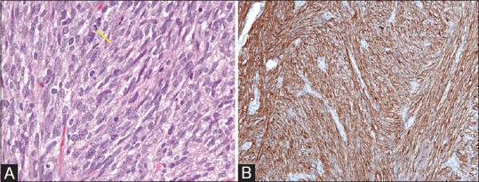
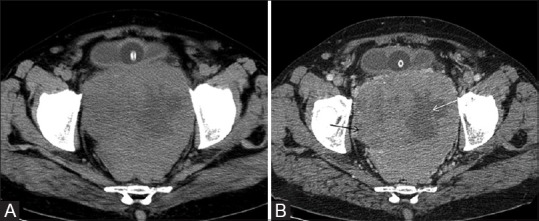
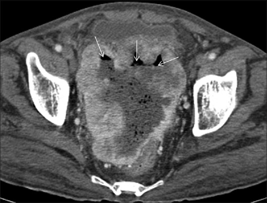
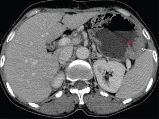
Gastrointestinal stromal tumor (GIST) are the most common non epithelial tumor of the gastrointestinal (GI) tract. They arise from interstitial cells of Cajal present in the myenteric plexus. They can also arise outside the GI tract from mesentery, retro peritoneum and omentum. With the advent of new targeted molecular therapy c- tyrosine kinase inhibitor (Imatinib), it has become important to differentiate between response and pseudo-progression of the disease as response evaluation criteria for GIST are different from Response Evaluation Criteria in Solid Tumors (RECIST). Purpose of this pictorial essay is to enumerate the characteristic CT features of GIST, and discuss atypical features and response evaluation criteria.

Keywords: GIST; gastrointestinal; imatinib; tumor.


Conflict of interest statement

There are no conflicts of interest.

Figures

Figure 1 (A and B)
Figure 1 (A and B)
(A) H and E, ×40; Cellular proliferation of spindle cells with pale to eosinophilic fibrillar cytoplasm, arranged in whorls or short intersecting fascicles. Rare mitotic activity (arrow) (B) IHC, ×40; Immunohistochemistry for CD117 (CKIT) shows strong and diffuse cytoplasmic staining
Figure 2 (A and B)
Figure 2 (A and B)
(A), pre contrast CT scan showing rectal GIST with heterogeneous attenuation with hypoattenuating areas within. (B) Post contrast CT scan heterogeneous GIST arising from the rectum with heterogeneous enhancement with non-enhancing cystic/necrotic areas within (arrows). It shows extraserosal extension anteriorly and abuts the urinary bladder without infiltration
Figure 3
Figure 3
Post contrast axial CT scan showing sigmoid GIST with peripheral post contrast enhancement. Central non enhancing cystic/necrotic area is seen. Air fluid levels (arrows) are also noted within suggestive of communication with gut
Figure 4
Figure 4
Axial post contrast CT scan showing homogeneously enhancing mass along the greater curvature of stomach (white arrow). Ulceration in the stomach is noted (red arrow)
Figure 5
Figure 5
There is hypoattenuating GIST involving the body of the stomach. There is central necrotic component communicating with the lumen of the stomach with resultant air in the tumor cavity (arrows)
Figure 6
Figure 6
Axial post contrast CT scan showing homogeneously enhancing exophytic gastric mass in the gastrohepatic region (white arrow) with few hypodense metastatic liver lesions (red arrow)
Figure 7 (A and B)
Figure 7 (A and B)
Ileal GIST with hyperdense area within (red arrow in Figure A) and shows no post contrast enhancement (red arrow in B) is hemorrhage within the tumor. Figure B shows heterogeneous enhancement in this tumor with non-enhancing hemorrhagic (red arrow) and necrotic/cystic components within (white arrows). Also there is heterogeneously enhancing liver metastasis (yellow arrow in B)
Figure 8 (A and B)
Figure 8 (A and B)
In this case of ileal GIST, axial post contrast CT scan shows (A) heterogeneously enhancing liver metastasis (white arrow) and (B) predominantly peripherally enhancing pelvic peritoneal deposits with central non enhancing cystic/necrotic area (red arrow)
Figure 9
Figure 9
Axial post contrast CT scan showing sigmoid GIST with peripheral enhancement and central non enhancing cystic/necrotic area. Specks of calcifications (hyperdense foci) are also seen within
Figure 10 (A and B)
Figure 10 (A and B)
Axial post contrast CT showing (A) rectal GIST with intraluminal extension and showing heterogeneous post contrast enhancement (white arrow) and (B) anal canal GIST with intraluminal extension (yellow arrow)
Figure 11 (A and B)
Figure 11 (A and B)
(A) Axial post contrast CT scan showing heterogeneously enhancing mesenteric GIST (white arrow) with central non enhancing cystic/necrotic area (yellow arrow). No communication to GI tract was seen. Histopathology showed features of GIST. (B) Axial post contrast CT scan showing homogeneously enhancing peritoneal GIST in left lumbar region (white arrow). No communication to GI tract was seen. Histopathology showed features of GIST
Figure 12
Figure 12
In this case of metastatic gastric GIST (image not shown), axial contrast enhanced CT scan shows heterogeneously enhancing masses in bilateral suprarenal regions involving both adrenal glands with non-enhancing cystic/necrotic areas (arrows), suggestive of bilateral adrenal metastases
Figure 13 (A and B)
Figure 13 (A and B)
In this case of gastric GIST, axial post contrast CT scan shows a large hypoattenuating metastatic liver lesion (white arrow in A). Post chemotherapy, there is decrease in size as well as attenuation of the liver metastatic lesion (yellow arrow in B). Residual gastric GIST is also seen (red arrow in B; not shown in Figure A)
Figure 14 (A and B)
Figure 14 (A and B)
(A) Axial post contrast CT image shows homogeneous wall thickening involving body of stomach (white arrow). Peripherally enhancing cystic/necrotic peritoneal deposit is also seen in left hypochondriac region (red arrow). Left pleural effusion is also seen (yellow arrow). (B) FDG PET/CT fused axial image shows intense FDG uptake in gastric GIST (white arrow). There is also peripheral FDG uptake in peritoneal deposit (red arrow). Central non FDG avid area is cystic/necrotic. (C) Post imatinib, there is no FDG uptake in gastric mass (white arrow) and interval decrease in size and FDG uptake (SUV less than 2.5) in peritoneal deposit (red arrow) suggestive response to imatinib

Similar articles

Cited by

References

    1. Lakshmi VA, Chacko RT, Kurian S. Gastrointestinal stromal tumors: A 7-year experience from a tertiary care hospital. Indian J Pathol Microbiol. 2010;53:628–33. - PubMed
    1. Joensuu H, Fletcher C, Dimitrijevic S, Silberman S, Roberts P, Demetri G. Management of malignant gastrointestinal stromal tumours. Lancet Oncol. 2002;3:655–64. - PubMed
    1. Rubin BP, Heinrich MC, Corless CL. Gastrointestinal stromal tumour. Lancet. 2007;19(369):1731–41. - PubMed
    1. Steigen SE, Eide TJ. Gastrointestinal stromal tumors (GISTs): A review. APMIS. 2009;12(117):73–86. - PubMed
    1. Nishida T, Hirota S. Biological and clinical review of stromal tumors in the gastrointestinal tract. Histol Histopathol. 2000;15:1293–301. - PubMed