. 2019 Apr-Jun;29(2):141-154.
doi: 10.4103/ijri.IJRI_465_18.

Barium esophagogram in various esophageal diseases: A pictorial essay

Affiliations

Barium esophagogram in various esophageal diseases: A pictorial essay

Uma Debi et al. Indian J Radiol Imaging. 2019 Apr-Jun.

Abstract

Recent years have seen a decline in number of barium procedures due to wider availability of cross sectional imaging modalities. Though use of barium esophagography/barium swallow has decreased in day to day clinical practice, it still remains a valuable test for structural and functional evaluation of esophagus. It can be performed as single or double contrast examination or as a multiphasic examination comprising upright double contrast views followed by prone single contrast views. This pictorial essay demonstrates imaging features of various esophageal diseases on barium esophagogram.

Keywords: Barium swallow; diverticula; esophagus; motility disorder; tumours; webs.


Conflict of interest statement

There are no conflicts of interest.

Figures

Figure 1 (A-C)
Figure 1 (A-C)
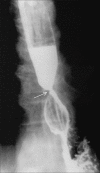
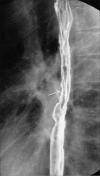
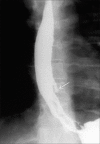
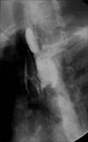
(A) Upright LPO view of single-contrast barium swallow demonstrates a shelf-like filling defect along the anterior wall of hypopharynx, at pharyngioesophageal junction s/o web. (B) Jet phenomenon associated with cervical web, usually seen in cases of partial obstruction. (C) Prominent cricopharyngeus muscle may mimic a web. However, it is seen along the posterior wall
Figure 2
Figure 2
Prone RAO view of single-contrast barium swallow demonstrating Schatzki ring
Figure 3 (A-C)
Figure 3 (A-C)
(A) Upright AP view of single-contrast barium swallow shows a pulsion diverticulum. Also, note multiple nonperistaltic contractions associated with motility dysfunction. (B) Upright AP view of single-contrast barium swallow showing a large epiphrenic diverticulum. (C) Upright lateral view of single-contrast barium swallow demonstrating a traction diverticulum arising from anterior wall of mid esophagus. Note the triangular appearance of traction diverticulum
Figure 4 (A-C)
Figure 4 (A-C)
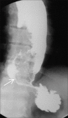
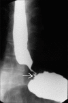
(A) Primary achlasia: Prone single-contrast barium swallow showing dilated esophagus with tapered beak-like narrowing at GE junction (arrow). On fluoroscopy, primary peristalsis in esophagus was absent. (B) Dilated and tortuous esophagus in a case of primary achlasia (C) Secondary achlasia: Prone RAO view showing mildly dilated esophagus with narrowing involving lower thoracic esophagus and GE junction, with mucosal irregularity (arrow). Note greater length of narrowed segment and less dilatation of esophagus in comparison to (A)
Figure 5
Figure 5
Diffuse esophageal spasm: upright single-contrast barium swallow image showing multiple nonperistaltic contractions in esophagus giving classic “corkscrew appearance”
Figure 6
Figure 6
Upright single-contrast barium swallow in a patient of scleroderma showed dilated esophagus with absent primary peristalsis in mid and lower esophagus. Also GE junction was patulous with presence of reflux and sliding hiatus hernia (differentiating it from achalasia)
Figure 7
Figure 7
Peptic stricture (arrow) in a patient with long standing history of gastroesophageal reflux. Upright double-contrast barium swallow shows sliding hiatus hernia with segment of smooth, concentric narrowing in lower esophagus. Also note proximal hold up of the barium
Figure 8
Figure 8
Barrett's esophagus: Upright LPO image of double-contrast barium swallow showing mild narrowing (arrow) and reticular pattern in columner epithelium at mid thoracic esophagus. Gastroesophageal reflux was also present in the same patient. Endoscopic biopsy revealed columnar metaplasia
Figure 9
Figure 9
HIV esophagitis: Upright single-contrast barium swallow image showing giant flat ulcer (arrow) arising from left posterolateral wall of mid esophagus in an HIV-positive patient
Figure 10
Figure 10
Stricture secondary to radiation: Case of squamous cell carcinoma of mid esophagus, postradiotherapy status. Barium swallow done 9 months after the completion of radiotherapy revealed long segment of concentric smooth narrowing involving distal esophagus
Figure 11 (A-C)
Figure 11 (A-C)
Caustic esophagitis: Upright frontal (A and B) and lateral (C) single contrast barium swallow images demonstrating long segment stricture involving distal esophagus in a patient with history of accidental lye ingestion. Also note presence of intramural pseudodiverticulae associated with the stricture (B)
Figure 12
Figure 12
Drug-induced esophagitis: Upright barium swallow image demonstrating ulceration (arrow) in mid esophagus with esophageal spasm. The esophagitis was related to intake of doxycycline
Figure 13
Figure 13
Leiomyoma: Upright double-contrast barium swallow image showing smooth crescentic filling defect in distal esophagus forming obtuse angle with esophageal wall (arrow) characteristic of a submucosal mass
Figure 14
Figure 14
Fibrovascular polyp: Upright barium swallow image showing expansion of cervical and proximal thoracic esopahgus with polyploidal filling defect (arrow)
Figure 15 (A-D)
Figure 15 (A-D)
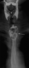
Esophageal carcinoma: (A) Early esophageal cancer seen as plaque-like lesion with mucosal irregularity and mild reduce distensibility (arrow) in mid thoracic esophagus on double-contrast barium swallow. (B) Infiltrative lesion: Prone RAO barium swallow image showing irregular infiltrative lesion (arrow) with shouldering in distal esophagus extending till GE junction. Also note proximal dilatation of esophagus. Endoscopic biopsy revealed the lesion to be adenocarcinoma. (C) Ulcerative lesion: Upright single-contrast barium swallow image demonstrating irregular ulcerative lesion in upper esophagus (arrow) without significant luminal narrowing. (D) Polypoidal lesion: Upright lateral view of single-contrast barium swallow showing polyploidal filling defect in upper esophagus (arrow). Patient had severe dysphagia and aspirated small amount of barium during the procedure
Figure 16
Figure 16
Spindle cell carcinoma: Upright frontal and LPO images of barium swallow demonstrating irregular polyploidal filling defect (arrow) in mid esophagus causing expansion of esophageal lumen
Figure 17
Figure 17
Leiomyosarcoma: Prone RAO double-contrast barium swallow image showing semilunar filling defect (arrow) in distal thoracic esophagus s/o submucosal mass. Endoscopic biopsy revealed leiomyosarcoma
Figure 18
Figure 18
Malignant melanoma: Upright frontal single-contrast barium swallow image showing irregular, large, polyploidal, expansile filling defect in distal esopahgus (arrow). This may be difficult to distinguish from spindle cell carcinoma
Figure 19
Figure 19
Non-Hodgkin's lymphoma of stomach involving GE junction: Concentric smooth narrowing involving distal esophagus (arrow). Multiple small polyploidal filling defects were also seen noted in stomach
Figure 20 (A and B)
Figure 20 (A and B)
(A) Sliding hiatus hernia: Upward migration of GE junction with herniation of stomach into thoracic cavity. (B) Mixed hernia: Upright barium swallow image showing paraesophageal herniation of fundus and body of stomach (arrow). Also note higher position of GE junction
Figure 21
Figure 21
Esophageal tuberculosis: Upright barium swallow image of a patient with history of dysphasia showing extrinsic impression in mid esophagus with presence of mild mucosal ulceration and displacement of esophagus towards right side (arrow)
Figure 22
Figure 22
Esophageal Crohn's disease: Upright barium swallow image showing stricture of mid and lower esophagus (arrow)
Figure 23
Figure 23
Mallory Weiss tear: Upright single-contrast barium swallow image showing intramural contrast extravasation from esophagus at level of GE junction (arrow), in a patient with history of violent episode of vomiting
Figure 24
Figure 24
Congenital short esophagus: LPO view in an infant showing short esophagus with intrathoracic stomach
Figure 25
Figure 25
FB sequelae: LAO view barium swallow image of a patient with past history of denture impaction showing stricture at proximal esophagus with restricted passage of barium. Also note H-shaped tracheoesophageal fistula with opacification of trachea

Similar articles

Cited by

References

    1. Rubesin SE. Structural abnormalities of the pharynx. In: Gore RM, Levine MS, editors. Textbook of Gastrointestinal Radiology. 3rd ed. Philadelphia, Pa: Saunders; 2008. pp. 271–308.
    1. Shauffer IA, Phillips HE, Sequeira J. The jet phenomenon a manifestation of esophageal web. AJR. 1977;129:747–8. - PubMed
    1. Taylor AI, Stewart ET, Dodds WJ. The eophageal jet phenomenon revisited. AJR. 1990;155:289–90. - PubMed
    1. Schatzki R. The lower esophageal ring: Long term follow-up of symptomatic and asymptomatic rings. Am J Roentgenol Ra-dium Ther Nucl Med. 1963;90:805–10. - PubMed
    1. Levine MS. Miscellaneous abnormalities of the esophagus. In: Gore RM, Levine MS, editors. Textbook of Gastrointestinal Radiology. 3rd ed. Philadelphia, Pa: Saunders; 2000. pp. 465–93.