. 2019 Apr-Jun;29(2):201-210.
doi: 10.4103/ijri.IJRI_484_18.

Imaging in chronic pancreatitis: State of the art review

Affiliations

Imaging in chronic pancreatitis: State of the art review

Rohan Kamat et al. Indian J Radiol Imaging. 2019 Apr-Jun.

Abstract

Chronic pancreatitis (CP) is an important gastrointestinal cause of morbidity worldwide. It can severely impair the quality of life besides life-threatening acute and long-term complications. Pain and pancreatic exocrine insufficiency (leading to malnutrition) impact the quality of life. Acute complications include pseudocysts, pancreatic ascites, and vascular complications. Long-term complications are diabetes mellitus and pancreatic cancer. Early diagnosis of CP is crucial to alter the natural course of the disease. However, majority of the cases are diagnosed in the advanced stage. The role of various imaging techniques in the diagnosis of CP is discussed in this review.

Keywords: Chronic pancreatitis; diagnosis; imaging.


Conflict of interest statement

There are no conflicts of interest.

Figures
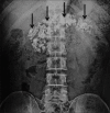
Figure 1
Figure 1
Plain abdominal radiograph shows extensive pancreatic calcifications (arrows)
Figure 2
Figure 2
Abdominal ultrasound shows atrophic pancreas with dilated main pancreatic duct (short arrow) with intraductal calculi (arrow)
Figure 3 (A and B)
Figure 3 (A and B)
EUS in a patient with chronic pancreatitis shows echogenic strands (arrow) in the head of pancreas (A). EUS in another patient with chronic pancreatitis (B) shows dilatation of main pancreatic duct (arrow) and side branches (short arrow)
Figure 4 (A-C)
Figure 4 (A-C)
Contrast-EUS in a patient with mass forming chronic pancreatitis shows a mass in the head of pancreas with cystic component (arrow, A) with peripheral enhancement (arrow, B) and central nonenhancing component. The central nonenhancing component is slightly echogenic than the rest of the mass (arrow, C). In long standing cases of mass forming chronic pancreatitis, this lack of enhancement mimics pancreatic adenocarcinoma
Figure 5 (A and B)
Figure 5 (A and B)
EUS elastography in a patient with chronic pancreatitis shows heterogenous stiffness of the pancreas (A) with hard areas (arrow) and areas of intermediate stiffness (short arrow). In another patient with mass forming chronic pancreatitis (B), elastography shows that the mass is hard (blue areas). Also note the presence of a cyst (arrow)
Figure 6 (A-F)
Figure 6 (A-F)
CT findings in chronic pancreatitis: Non-contrast CT (A) shows extensive pancreatic calcifications (arrows). CT scan in another patient (B) shows pancreatic atrophy (arrow) and a small cystic area (short arrow). Pancreatic duct irregularity and varying degrees of dilatation is shown in C to E (arrow). A large pseudocyst is also seen in d. An intraductal calculus (arrow) with pancreatic head mass is seen in another patient (F)
Figure 7 (A-E)
Figure 7 (A-E)
Diffusion weighted MRI (DWI) in normal patient and patient with chronic pancreatitis: Axial T1-weighted image (A) in a normal subject shows diffuse hyperintensity of the pancreas (arrow). The corresponding DWI (B) shows no diffusion restriction (arrow). Axial T1-weighted image in a patient with focal autoimmune pancreatitis (C) shows a hypointense lesion in tail of pancreas. On corresponding DWI (D) and ADC (E), there is evidence of diffusion restriction (arrow)
Figure 8 (A and B)
Figure 8 (A and B)
MRI findings in chronic pancreatitis: Axial T1-weighted contrast enhanced MRI (A) shows reduced T1-weighted signal of the pancreas (arrow). Axial T1-weighted contrast enhanced MRI in another patient (B) shows mild reduction in bulk with a cystic lesion in neck of pancreas (arrow)
Figure 9 (A-C)
Figure 9 (A-C)
MRCP findings in chronic pancreatitis: Multiple strictures (arrow) as well as filling defects (short arrow) within the main pancreatic duct are seen in A. In another patient (B), a large intraductal calculus (arrow) is causing marked dilatation of the pancreatic duct. MRCP in a different patient (C) shows strictures (arrow) and irregularity (short arrow) of the pancreatic duct
Figure 10 (A-C)
Figure 10 (A-C)
ERCP findings in chronic pancreatitis: In a patient with early chronic pancreatitis (A), mild dilatation of the main pancreatic duct (arrow) and side branches (short arrow) is seen. Marked ductal dilatation (arrow) with a dominant stricture (short arrow) is shown in B. In another patient (C), strictures (arrow) and intraductal calculi (short arrow) are seen
Figure 11 (A-D)
Figure 11 (A-D)
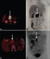
FDG-PET in chronic pancreatitis: Axial PET image (A) and corresponding coronal MIP image (B) shows marked FDG avidity (SUV max-12) in the pancreatic head mass in a patient with pancreatic adenocarcinoma. In a patient with mass forming chronic pancreatitis show PET shows low FDG avidity in the pancreatic head mass (arrow, C and D)
Figure 12 (A-D)
Figure 12 (A-D)
Imaging features in autoimmune pancreatitis: Axial CT image (A) shows sausage shaped pancreas (arrow). In a different patient, CT (B) shows sausage shaped pancreas (arrow) with peripheral hypodense rim (short arrow). MRI in the same patient (C) shows slightly bulky sausage shaped pancreas (arrow) with hypointense rim (short arrow). MRCP in another patient (D) with autoimmune pancreatitis shows hilar stricture (arrow)
Figure 13
Figure 13
Focal autoimmune pancreatitis: EUS demonstrated a mass in the head of pancreas (arrow, A) with predominantly hard areas (blue areas, arrow, B)
Figure 14
Figure 14
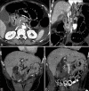
CT findings in groove pancreatitis: Axial (A) and coronal (B) CT images show thickening of the duodenum (arrow, A) with a hypodense soft tissue in the pancreatoduodenal groove (arrow, B). Coronal CT images (C and D) in another patient show a more pronounced hypodense soft tissue in the pancreatoduodenal groove (arrows). Also note the dilatation of the pancreatic duct (short arrow, C)
Figure 15
Figure 15
EUS in groove pancreatitis: Thickening of the duodenal wall with a cyst (arrow, A) is seen. The tissue in the pancreatoduodenal groove is hard on elastography (arrow B, blue and green areas)

Similar articles

Cited by

References

    1. De Backer AI, Mortele KJ, Ros RR, Vanbeckevoort D, Vanschoubroeck I, De Keulenaer B. Chronic pancreatitis: Diagnostic role of computed tomography and magnetic resonance imaging. JBR-BTR. 2002;85:304–10. - PubMed
    1. Sharma R, Kandasamy D. Imaging in chronic pancreatitis. Pancreapedia Exocrine Pancreas Knowl Base. 2015
    1. Perez-Johnston R, Sainani NI, Sahani DV. Imaging of chronic pancreatitis (including groove and autoimmune pancreatitis) Radiol Clin North Am. 2012;50:447–66. - PubMed
    1. Busireddy KK, AlObaidy M, Ramalho M, Kalubowila J, Baodong L, Santagostino I, et al. Pancreatitis-imaging approach. World J Gastrointest Pathophysiol. 2014;5:252–70. - PMC - PubMed
    1. Siddiqi AJ, Miller F. Chronic pancreatitis: Ultrasound, computed tomography, and magnetic resonance imaging features. Semin Ultrasound CT MRI. 2007;28:384–94. - PubMed