

Role of interventional radiology in the diagnosis and management of congenital extrahepatic portosystemic shunts: Two case reports
- PMID: 31367096
- PMCID: PMC6639860
- DOI: 10.4103/ijri.IJRI_461_18
Role of interventional radiology in the diagnosis and management of congenital extrahepatic portosystemic shunts: Two case reports
Abstract
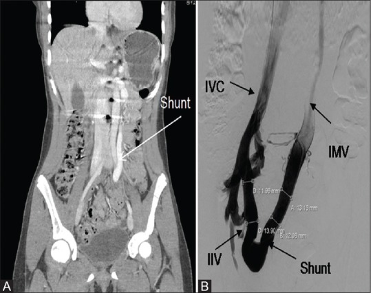
Congenital extrahepatic portosystemic shunt (CEPS) is a rare splanchnic venous malformation, wherein the portal venous outflow drains into the systemic venous circulation via a pathologic shunt. CEPS exhibits heterogeneous clinical behavior and angiography is the gold standard for evaluation of the portomesenteric communication to systemic vasculature. The potential severity of complications necessitates shunt closure. Here, we present two cases of CEPS. The first patient presented with an asymptomatic hyperammonemia and was found to have a Type 1 CEPS with absence of intrahepatic portal system. The second patient was asymptomatic and was incidentally found to have a Type 2 CEPS on imaging with normal intrahepatic portal system. Both patients were successfully treated with endovascular occlusion of the CEPS.
Keywords: Congenital extrahepatic portosystemic shunt; endovascular closure; focal nodular hyperplasia; hyperammonemia; portal hypertension.
Conflict of interest statement
There are no conflicts of interest.
Figures


Similar articles
-
Congenital extrahepatic portosystemic shunt: an underdiagnosed but treatable cause of hepatopulmonary syndrome.Eur J Pediatr. 2016 Feb;175(2):195-201. doi: 10.1007/s00431-015-2623-4. Epub 2015 Aug 27. Eur J Pediatr. 2016. PMID: 26311567
-
Multistage closure of a congenital extrahepatic portosystemic shunt.CVIR Endovasc. 2021 Nov 18;4(1):79. doi: 10.1186/s42155-021-00267-x. CVIR Endovasc. 2021. PMID: 34792654 Free PMC article.
-
Role of ultrasonography in early diagnosis of congenital extrahepatic portosystemic shunt.BJR Case Rep. 2016 May 25;2(2):20150266. doi: 10.1259/bjrcr.20150266. eCollection 2016. BJR Case Rep. 2016. PMID: 30363630 Free PMC article.
-
Congenital extrahepatic portosystemic shunts.Pediatr Radiol. 2003 Sep;33(9):614-20. doi: 10.1007/s00247-003-1002-x. Epub 2003 Jul 23. Pediatr Radiol. 2003. PMID: 12879313 Review.
-
Clinical and radiologic manifestations of congenital extrahepatic portosystemic shunts: a comprehensive review.Radiographics. 2011 May-Jun;31(3):707-22. doi: 10.1148/rg.313105070. Radiographics. 2011. PMID: 21571652 Review.
Cited by
-
A basic understanding of congenital extrahepatic portosystemic shunt: incidence, mechanism, complications, diagnosis, and treatment.Intractable Rare Dis Res. 2020 May;9(2):64-70. doi: 10.5582/irdr.2020.03005. Intractable Rare Dis Res. 2020. PMID: 32494552 Free PMC article. Review.
-
Efficacy and Safety of Surgical Ligation versus Endovascular Embolization for Type II Congenital Extrahepatic Portosystemic Shunt.Biomed Res Int. 2021 May 31;2021:9951393. doi: 10.1155/2021/9951393. eCollection 2021. Biomed Res Int. 2021. PMID: 34159206 Free PMC article.
References
-
- Bernard O, Franchi-Abella S, Branchereau S, Pariente D, Gauthier F, Jacquemin E. Congenital portosystemic shunts in children: Recognition, evaluation, and management. Semin Liver Dis. 2012;32:273–87. - PubMed
-
- Alonso-Gamarra E, Parrón M, Pérez A, Prieto C, Hierro L, López-Santamaría M. Clinical and radiologic manifestations of congenital extrahepatic portosystemic shunts: A comprehensive review. Radiographics. 2011;31:707–72. - PubMed
-
- Murray CP, Yoo SJ, Babyn PS. Congenital extrahepatic portosystemic shunts. Pediatr Radiol. 2003;33:614–20. - PubMed