Meta-Analysis
. 2019 Jul 22:14:37.
doi: 10.1186/s13017-019-0254-1. eCollection 2019.

Pregnancy and appendicitis: a systematic review and meta-analysis on the clinical use of MRI in diagnosis of appendicitis in pregnant women

Affiliations
Meta-Analysis

Pregnancy and appendicitis: a systematic review and meta-analysis on the clinical use of MRI in diagnosis of appendicitis in pregnant women

Mania Kave et al. World J Emerg Surg. .

Abstract

Background: The aim of this systematic review and meta-analysis was to evaluate the clinical use of MRI for the evaluation of acute appendicitis during pregnancy.

Methods: The searches were conducted by two independent researchers (MK, MS) to find the relevant studies published from 1/1/2009 until end of 30/12/2018. We searched for published literature in the English language in MEDLINE via PubMed, EMBASETM via Ovid, The Cochrane Library, and Trip database. For literature published in other languages, we searched national databases (Magiran and SID), KoreaMed, and LILACS. The keywords used in the search strategy are Pregnancy [MeSH], Pregnant [MeSH] OR-Magnetic resonance imaging [MeSH] OR-Appendicitis [MeSH] OR-Ultrasound, [MeSH] OR, imaging, MRI [MeSH] OR"،" and Right lower quadrant pain [MeSH]. The risk of bias of every article was evaluated by using QUADAS-2. On the basis of the results from the 2 × 2 tables, pooled measures for sensitivity, specificity, diagnostic odds ratio (DOR), and area under the curves (AUC) along with their 95% confidence intervals (CIs) were calculated using the DerSimonian Lair methodology.

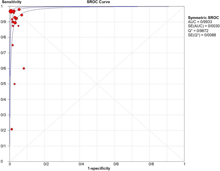
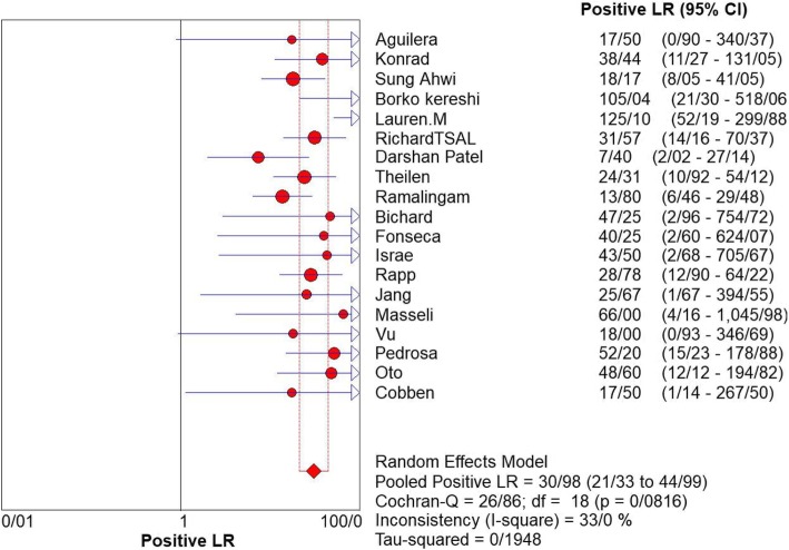
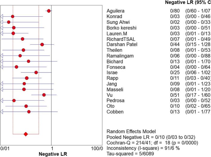
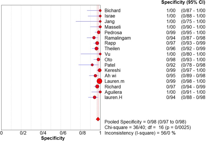
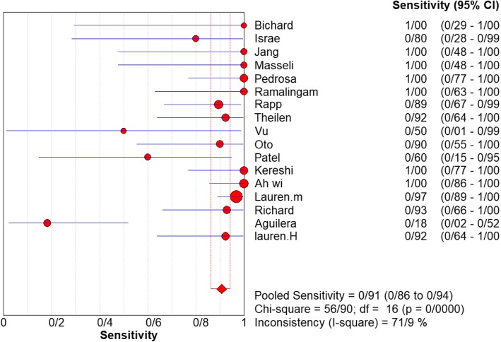
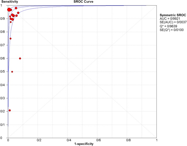
Results: As many as 1164 studies were selected. After analyzing the correspondence of the studies with the required criteria, 19 studies were selected for the final review. For appendicitis in pregnancy, the MRI sensitivity was 91.8% at the 95% confidence interval of (95% CI 87.7-94.9%). At the confidence interval of 95%, the specificity was 97.9% (95% CI 0.97.2-100%). The risk of bias in the studies conducted was measured using the QUADAS-2 tool.

Conclusion: MRI has high sensitivity and specificity (91.8%, 97.9% respectively) for the diagnosis of acute appendicitis in pregnant patients with clinically suspected appendicitis. It is an excellent imaging technique in many instances, which does not expose a fetus, or the mother, to ionizing radiation, making it an excellent option for pregnant patients with suspected acute appendicitis.

Keywords: Acute appendicitis; Magnetic resonance imaging (MRI); Pregnancy.


Conflict of interest statement

Competing interestsThe authors declare that they have no competing interests.

Figures

Fig. 1
Fig. 1
PRISMA flow diagram
Fig. 2
Fig. 2
The risk of bias in the studies conducted was measured by using the QUADAS-2 tool. The risk of bias shown in Eq. 2 of the above image model (MRI) of each diagram indicate the number and percentage of studies with high (red), medium (yellow), and low (green) risk of bias in the four groups of the QUADAS-2 tool
Fig. 3
Fig. 3
Sensitivity of MRI for diagnosing appendicitis in studies that included pregnant patients only. Forest plot of sensitivity reported in each study. Each study is identified by name of first author and year of publication, with circles representing individual study point estimates, size of each circle indicating relative contribution to data pooling (inverse variance weighting), horizontal lines indicating 95% CIs, and dashed vertical lines representing 95% CIs for pooled sensitivity and specificity
Fig. 4
Fig. 4
Specificity of MRI (≥ 1.5 T) for diagnosing appendicitis in studies that included pregnant patients only. Forest plots of specificity reported in each study. Each study is identified by name of first author and year of publication, with circles representing individual study point estimates, size of each circle indicating relative contribution to data pooling (inverse variance weighting), horizontal lines indicating 95% CIs, and dashed vertical lines representing 95% CIs for pooled specificity
Fig. 5
Fig. 5
Summary-ROC (SROC) curve for diagnostic accuracy of MRI in diagnosing appendicitis. Size of each circle on graph represents sample size of included study. SE = standard error; Q* index = point at which sensitivity and specificity are equal or point closest to ideal top-left corner of SROC space
Fig. 6
Fig. 6
Positive LR of MRI for diagnosing appendicitis in studies that included pregnant patients only. Forest plot of positive LR reported in each study. Each study is identified by name of first author and year of publication, with circles representing individual study point estimates, size of each circle indicating relative contribution to data pooling (inverse variance weighting), horizontal lines indicating 95% CIs, and dashed vertical lines representing 95% CIs for pooled positive LR
Fig. 7
Fig. 7
Negative LR of MRI for diagnosing appendicitis in studies that included pregnant patients only. Forest plot of negative LR reported in each study. Each study is identified by name of first author and year of publication, with circles representing individual study point estimates, size of each circle indicating relative contribution to data pooling (inverse variance weighting), horizontal lines indicating 95% CIs, and dashed vertical lines representing 95% CIs for pooled Negative LR
Fig. 8
Fig. 8
Specifity of MRI (≥ 1.5 T) for diagnosing appendicitis in studies that included pregnant patients only. Forest plot of sensitivity reported in each study. Each study is identified by name of first author and year of publication, with circles representing individual study point estimates, size of each circle indicating relative contribution to data pooling (inverse variance weighting), horizontal lines indicating 95% CIs, and dashed vertical lines representing 95% CIs for pooled sensitivity and specificity
Fig. 9
Fig. 9
Sensivity of MRI (≥ 1.5 T) for diagnosing appendicitis in studies that included pregnant patients only. Forest plots of specificity reported in each study. Each study is identified by name of first author and year of publication, with circles representing individual study point estimates, size of each circle indicating relative contribution to data pooling (inverse variance weighting), horizontal lines indicating 95% CIs, and dashed vertical lines representing 95% CIs for pooled specificity
Fig. 10
Fig. 10
Summary-ROC (SROC) curve for diagnostic accuracy of MRI (≥ 1.5 T) in diagnosing appendicitis. Size of each circle on graph represents sample size of included study. SE = standard error; Q* index = point at which sensitivity and specificity are equal or point closest to ideal top-left corner of SROC space
Fig. 11
Fig. 11
Pooled sensitivity and specificity of MRI in diagnosis of appendicitis in pregnancy by countries

References

    1. Ueberrueck T, Koch A, Meyer L, et al. Ninety-four appendectomies for suspected acute appendicitis during pregnancy. World J Surg. 2004;28(5):508–511. doi: 10.1007/s00268-004-7157-2. - DOI - PubMed
    1. Andersson Roland EB, Lambe Mats. Incidence of appendicitis during pregnancy. International Journal of Epidemiology. 2001;30(6):1281–1285. doi: 10.1093/ije/30.6.1281. - DOI - PubMed
    1. Şimşek Deniz, Turan Özgür Deniz, Ergenoğlu Ahmet Mete, Demir Halit Batuhan, Sezer Taylan Özgür, Şahin Çağdaş. Pregnancy Outcomes and Surgical Management of Pregnancy Complicated By Appendicitis: Obstetrician View. Meandros Medical and Dental Journal. 2015;16(2):43–49. doi: 10.4274/meandros.2401. - DOI
    1. Apandisit AP. Acute perforated appendicitis as a cause of fetal tachycardia at term pregnancy. Cukurova Med J. 2015;40(2):336–339. doi: 10.17826/cutf.83967. - DOI
    1. Aydın S, Fatihoğlu E. Perfore Apandisit: Ultrasonografik Bir Tanısal Zorluk. Ankara Eğitim ve Araştırma Hastanesi Tıp Dergisi. 51(2):110–5.