. 2019 Jun 24;9(16):4633-4647.
doi: 10.7150/thno.32260. eCollection 2019.

Human Mesenchymal Stem Cell-Treated Regulatory CD23+CD43+ B Cells Alleviate Intestinal Inflammation

Affiliations

Human Mesenchymal Stem Cell-Treated Regulatory CD23+CD43+ B Cells Alleviate Intestinal Inflammation

Xiaoyong Chen et al. Theranostics. .

Abstract

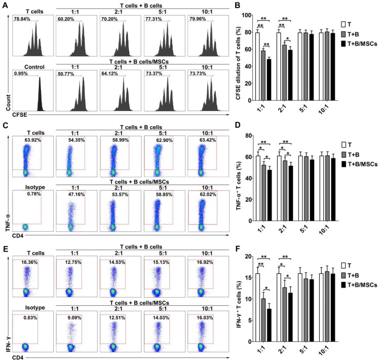
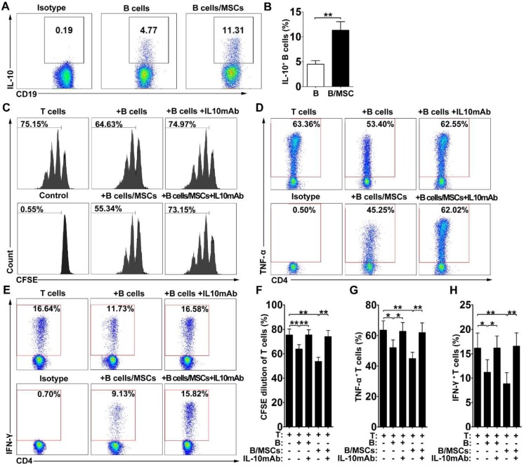
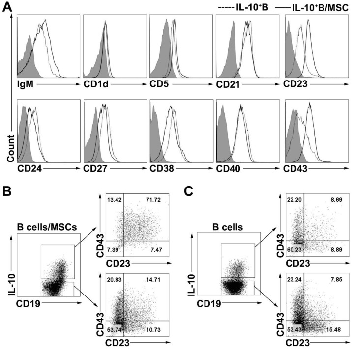
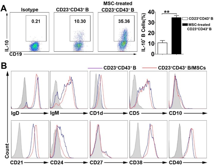
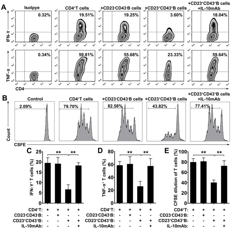
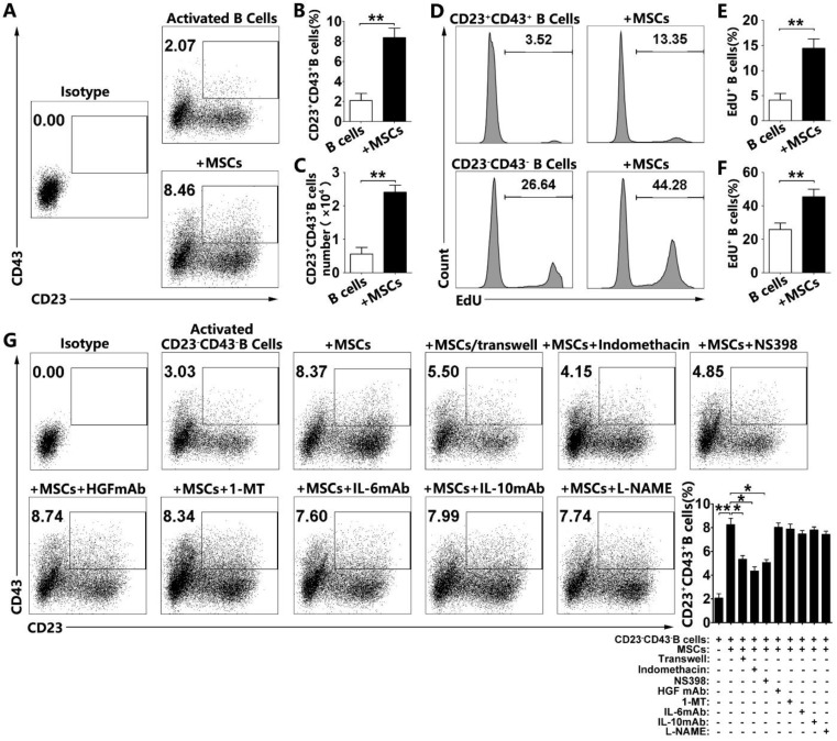
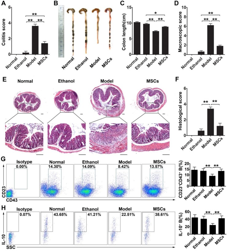
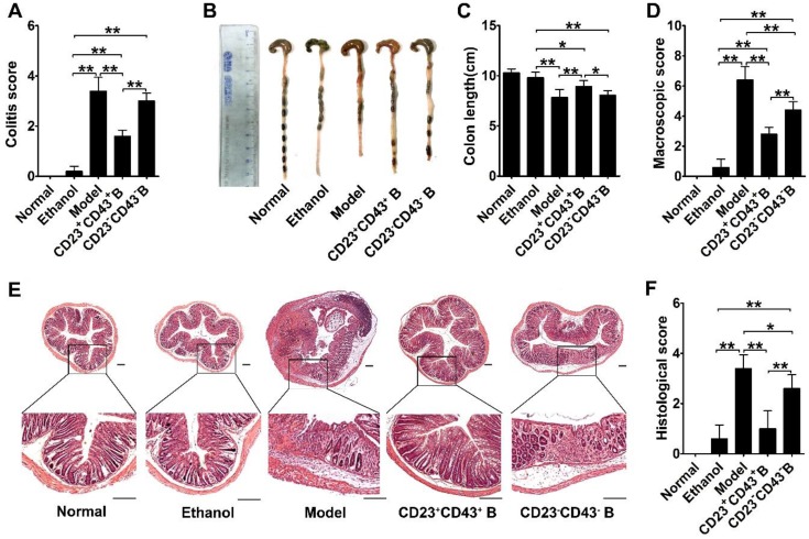
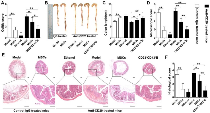
Rationale: Mesenchymal stem cells (MSCs) have been demonstrated to ameliorate inflammatory bowel disease by their actions on multiple immune cells, especially on regulatory B cells (Breg cells). However, the phenotypes and functions of human MSCs (hMSCs)-treated Breg cell subsets are not yet clear. Methods: Purified B cells were cocultured with MSCs and the phenotypes and immunomodulatory functions of the B cells were analyzed by FACS and proliferation assays in vitro. Also, a trinitrobenzenesulfonic acid-induced mouse colitis model was employed to detect the function of MSC-treated Breg cells in vivo. Results: We demonstrated that coculturing with hMSCs significantly enhanced the immunomodulatory activity of B cells by up-regulating IL-10 expression. We then identified that a novel regulatory B cell population characterized by CD23 and CD43 phenotypic markers could be induced by hMSCs. The CD23+CD43+ Breg cells substantially inhibited the inflammatory cytokine secretion and proliferation of T cells through an IL-10-dependent pathway. More significantly, intraperitoneal injection of hMSCs ameliorated the clinical and histopathological severity in the mouse experimental colitis model, accompanied by an increase in the number of CD23+CD43+ Breg cells. The adoptive transfer of CD23+CD43+ B cells effectively alleviated murine colitis, as compared with the CD23-CD43- B cells. Treatment with CD23+CD43+ B cells, and not hMSCs, substantially improved the symptoms of colitis in B cell-depleted mice. Conclusion: the novel CD23+CD43+ Breg cell subset appears to be involved in the immunomodulatory function of hMSCs and sheds new light on elucidating the therapeutic mechanism of hMSCs for the treatment of inflammation-related diseases.

Keywords: inflammatory bowel disease; interleukin-10; mesenchymal stem cells; regulatory B cells.


Conflict of interest statement

Competing Interests: The authors have declared that no competing interest exists.

Figures

Figure 1
Figure 1
hMSCs enhanced the immunosuppressive effect of B cells on T cell responses. CpG ODN 2006 and CD40L-stimulated B cells or hMSCs-treated B cells were cocultured with sorted CD4+ T cells, and the proliferation and pro-inflammatory cytokine secretion of T cells were evaluated by flow cytometry. (A) Representative histograms of CFSE-labeled T cell proliferation at different T/B ratios. (B) Quantification of CFSE-diluted T cells. (C) Representative plots of TNF-α production by T cells at different T/B ratio. (D) Quantification of TNF-α-producing T cells. (E) Representative plots of IFN-γ production by T cells in different T/B ratio. (F) Quantification of IFN-γ-producing T cells. Data represent mean values ± SD of four independent experiments. *p<0.05; **p<0.01.
Figure 2
Figure 2
hMSCs enhanced the immunosuppressive effect of B cells by inducing Breg cells. B cells were cultured alone or with hMSCs in the presence of CpG ODN 2006 and CD40L for 48h, and the percentage of IL-10 producing B cells were assessed. (A) Representative plots of IL-10 production by B cells with or without hMSCs. (B) Quantification of IL-10-producing B cells. The anti-IL-10 neutralizing antibodies were added to the T/B coculture, and the alteration of T cell responses was evaluated. (C) Representative histograms of CFSE-labeled T cells' proliferation with or without IL-10 blocking. (D) Representative plots of TNF-α production by T cells with or without IL-10 blocking. (E) Representative plots of IFN-γ production by T cells with or without IL-10 blocking. (F) Quantification of CFSE-diluted T cells. (G) Quantification of TNF-α-producing T cells. (H) Quantification of IFN-γ-producing T cells. Data represent mean values ± SD of four independent experiments. *p<0.05; **p<0.01.
Figure 3
Figure 3
hMSCs induced IL-10-producing B cells highly expressed CD23 and CD43. B cells were cultured alone or with hMSCs in the presence of CpG ODN 2006 and CD40L for 48h, and the surface markers of B cells were detected. (A) Representative cell surface phenotype of B10 cells and hMSC-educated B10 cells. Filled histograms indicate isotype controls, the solid line represents the hMSC-educated IL-10+ B cells, and the dotted line represents the IL-10+ B cells without hMSCs. (B) Representative dot plots of CD23 and CD43 expression on IL10+ and IL10- B cells cultured with hMSCs. (C) Representative dot plots of CD23 and CD43 expression on IL10+ and IL10- B cells without hMSCs.
Figure 4
Figure 4
The characteristics of hMSC-induced CD23+CD43+ B cells. B cells were cultured with hMSCs in the presence of CpG ODN 2006 and CD40L for 48h, and the IL-10 producing ability and surface markers of hMSC-induced CD23+CD43+ B cells were detected. (A) Representative plots of IL-10 production by CD23+CD43+ B cells with or without hMSCs. (B) Representative cell surface phenotype of CD23+CD43+ B cells with or without hMSCs. Filled histograms indicate isotype controls, blue lines represent CD23+CD43+ B cells without hMSCs, and red lines represent hMSCs-educating CD23+CD43+ B cells. Data represent mean values ± SD of five independent experiments. *p<0.05; **p<0.01.
Figure 5
Figure 5
CD23+CD43+ B cells inhibited the T cell response dependent on IL-10 production. B cells were cultured with hMSCs in the presence of CpG ODN 2006 and CD40L for 48h, then CD23+CD43+ B cells and CD23-CD43- B cells were isolated from hMSC-educated B cells and cultured with T cells. (A) Representative plots of cytokine secretion. CD23+CD43+ B cells, but not CD23-CD43- B cells, suppressed the TNF-α and IFN-γ production by T cells that was reversed by anti-IL-10mAb. (B) Representative histograms of T cell proliferation. CD23+CD43+ B cells, but not CD23-CD43- B cells, suppressed the proliferation of CFSE-labeled T cells that was reversed by anti-IL-10mAb. (C) Quantification of IFN-γ-producing T cells. (D) Quantification of TNF-α-producing T cells. (E) Quantification of CFSE-diluted T cells. Data represent mean values ± SD of four independent experiments. *p<0.05; **p<0.01.
Figure 6
Figure 6
hMSCs induced CD23+CD43+ B cells by promoting their proliferation and conversion from CD23-CD43- B cells by cell-to-cell contact and COX2/PGE2 pathway. Purified CD19+B cells or CD19+CD23-CD43- B cells were cultured alone or with hMSCs in the presence of CpG ODN 2006 and CD40L for 48h. (A) Representative plots of alteration in percentages of CD23+CD43+B cells in B cells with or without hMSCs. (B) Quantification of CD23+CD43+ B cell numbers. (C) Quantification of CD23+CD43+ B cell numbers. (D) Representative histogram of the proliferation of CD23+CD43+ and CD23-CD43- B cells alone or with hMSCs. (E) Quantification of the proliferation of CD23+CD43+ B cells. (F) Quantification of the proliferation of CD23-CD43- B cells. (G) Representative plots of increase in percentages of CD23+CD43+ B cells generated from CD23-CD43- B cells, quantification of CD23+CD43+ B cells. Data represent mean values ± SD of five independent experiments. *p<0.05; **p<0.01.
Figure 7
Figure 7
hMSCs alleviated colon inflammation in TNBS-induced colitis and CD23+CD43+ B cells recovery in the peritoneal cavity. Colitis was induced by TNBS in mice, which received the intraperitoneal injection of hMSCs or saline. Mice were euthanized on day 3 after treatment. (A) Colitis score of mice. (B) Representative colonic length of mice. (C) Quantification of the colonic length of mice. (D) Macroscopic damage score of colons. (E) Representative histological changes of colons. Scale bar=200 μm. (F) Histological score of colons. (G) Alteration in percentages of CD23+CD43+ B cells in the peritoneal cavity. (H) Changes in IL-10 production of CD23+CD43+ B cells in the peritoneal cavity. Data represent mean values ± SD of five mice per group. *p<0.05; **p<0.01.
Figure 8
Figure 8
CD23+CD43+ B cells adoptive transfer greatly reduced the severity of colitis. Colitis was induced by TNBS in mice by intraperitoneal injection of purified CD23+CD43+ B cells or CD23-CD43- B cells. Mice were euthanized on day 3 after treatment. (A) Colitis score of mice. (B) Representative colonic length of mice. (C) Quantification of the colonic length of mice. (D) Macroscopic damage score of colons. (E) Representative histological changes of colons. Scale bar=200 μm. (F) Histological score of colons. Data represent mean values ± SD of five mice per group. *p<0.05; **p<0.01.
Figure 9
Figure 9
CD23+CD43+ B cells but not hMSCs significantly mitigated colitis in B cell-depleted mice. Colitis was induced by TNBS in B cell-depleted mice, which received the intraperitoneal injection of hMSCs, CD23+CD43+ B cells, or saline. Mice were euthanized on day 3 after treatment. (A) Colitis score of mice. (B) Representative colonic length of mice. (C) Quantification of the colonic length of mice. (D) Macroscopic damage score of colons. (E) Representative histological changes of colons. Scale bar=200 μm. (F) Histological score of colons. Data represent mean values ± SD of five mice per group. *p<0.05; **p<0.01.

Similar articles

Cited by

References

    1. de Souza HS, Fiocchi C. Immunopathogenesis of IBD: current state of the art. Nat Rev Gastroenterol Hepatol. 2016;13:13–27. - PubMed
    1. Stappenbeck TS, Miyoshi H. The Role of Stromal Stem Cells in Tissue Regeneration and Wound Repair. Science. 2009;324:1666–9. - PubMed
    1. Duran NE, Hommes DW. Stem cell-based therapies in inflammatory bowel disease: promises and pitfalls. Therap Adv Gastroenterol. 2016;9:533–47. - PMC - PubMed
    1. Salem HK, Thiemermann C. Mesenchymal stromal cells: current understanding and clinical status. Stem cells. 2010;28:585–96. - PMC - PubMed
    1. Barzilay R, Melamed E, Offen D. Introducing transcription factors to multipotent mesenchymal stem cells: making transdifferentiation possible. Stem cells. 2009;27:2509–15. - PubMed

Publication types

MeSH terms