

Case Reports
doi: 10.1016/j.radcr.2019.07.003.
eCollection 2019 Sep.
Fetus in fetu: A rare case of intra-abdominal mass
Affiliations
- PMID: 31367265
- PMCID: PMC6656986
- DOI: 10.1016/j.radcr.2019.07.003
Item in Clipboard
Case Reports
Fetus in fetu: A rare case of intra-abdominal mass
Radiol Case Rep.
.
Abstract
Fetus in fetu is uncommon cause of retroperitoneal mass in infancy that resulted from abnormal embryogenesis. Clinical manifestations vary and mostly presented at infancy It is differentiated from teratoma through its location, benign course, and identification of limb buds and well-organized organs. Radiologically, identification of long bones and organized vertebral bodies are core in the diagnosis. Presented is 4 days-old male with abdominal distention as the main manifestation. Definite radiological diagnosis was done by Ultrasound (US) and computed tomography scan.
Keywords: Abdominal mass; Fetus in fetu; Teratoma.
Figures
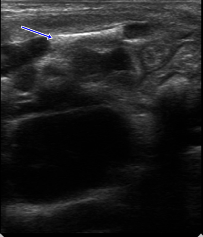
U/S: Shows a well-defined complex abdominal mass lesion with cystic and bony (Blue arrow) elements. (Color version of figure is available online.)
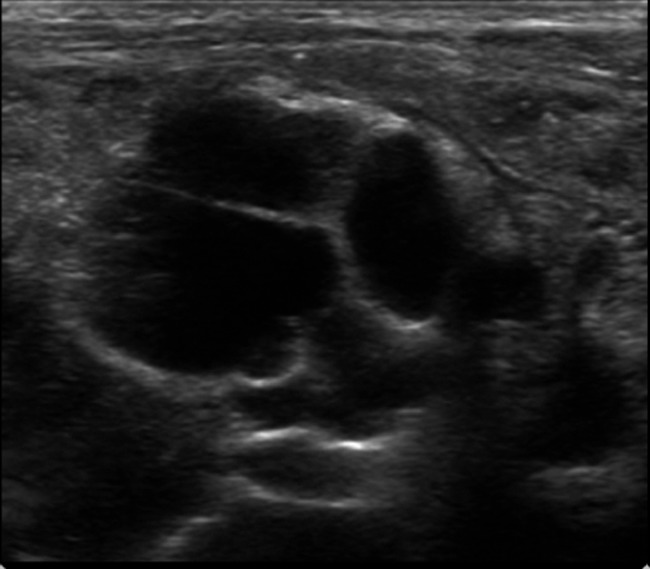
U/S shows a well-defined complex abdominal mass lesion with cystic elements.
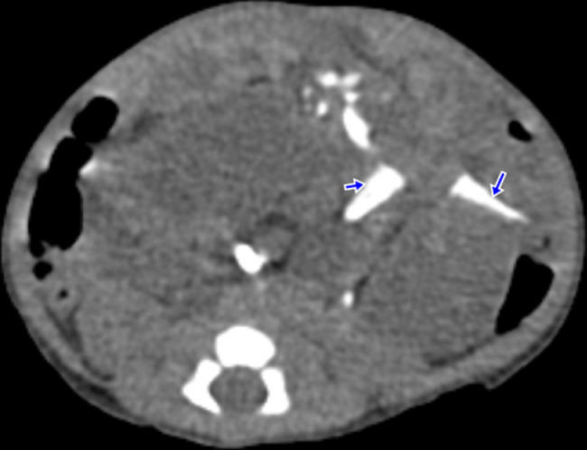
Axial plain CT showing an intra-abdominal mass lesion displacing the bowel loops and shows remnants of bone tissue (Blue arrows). (Color version of figure is available online.)
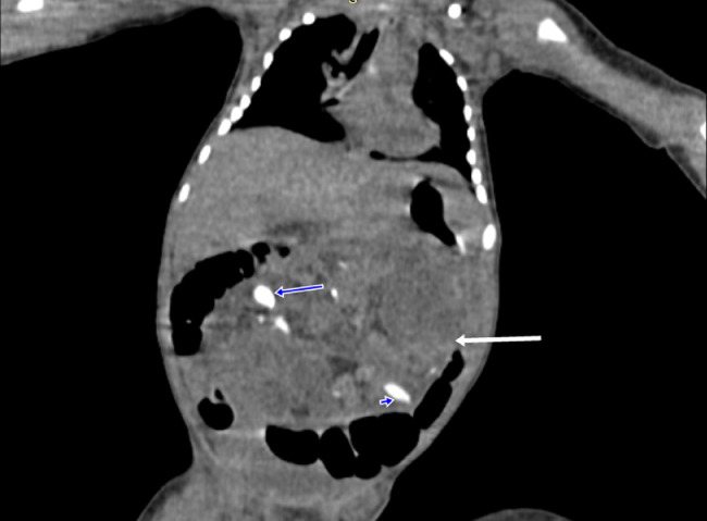
Coronal plain CT showing an intra-abdominal mass lesion (White arrow) displacing the bowel loops and shows remnants of bone tissue (Blue arrows). (Color version of figure is available online.)
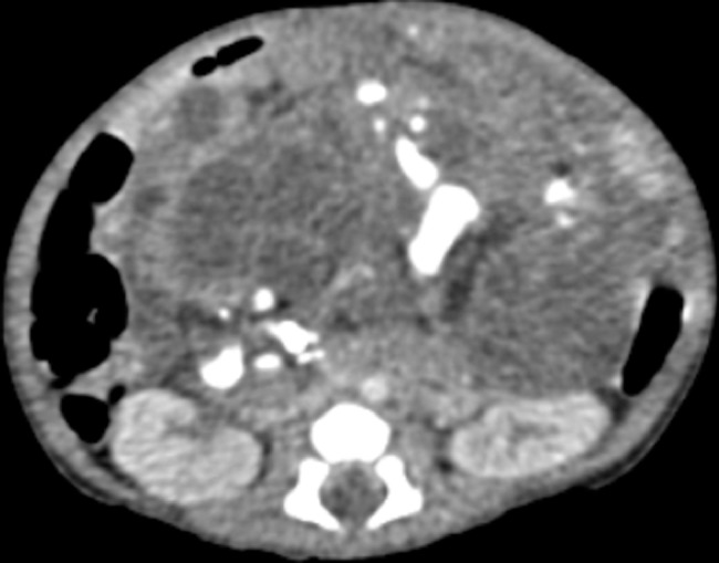
Axial CT with IV contrast showing a heterogenous intra-abdominal mass lesion displacing the bowel loops and shows remnants of bone tissue.
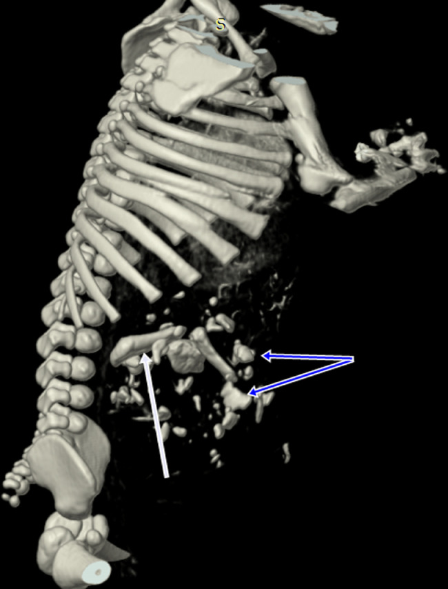
3D reconstruction of Fetus in Fetu showing long bones (Blue arrows) and dysmorphic vertebrae (White arrows). (Color version of figure is available online.)
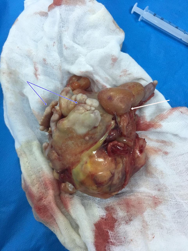
Excision of the mass through abdominal exploration under general anesthesia showing limb buds (Blue arrows) and the cystic components (White arrow).
References
-
- Arlikar J.D., Mane S.B., Dhende N.P., Sanghavi Y., Valand A.G., Butale P.R. Fetus in fetu: two case reports and review of literature. Pediatr Surg Int. 2009;25(3):289. - PubMed
-
- Willis R.A. The borderland of embryology and pathology. Bull N Y Acad Med. 1950;26(7):440–460. https://www.ncbi.nlm.nih.gov/pubmed/15426876 Available from: - PMC - PubMed
-
- Patankar T., Fatterpekar G.M., Prasad S., Maniyar A., Mukherji S.K. Fetus in fetu: CT appearance—report of two cases. Radiology. 2000;214(3):735–737. - PubMed
Publication types
LinkOut - more resources
Full Text Sources
