Review
. 2019 May 23;10(26):6404-6422.
doi: 10.1039/c9sc01545k. eCollection 2019 Jul 14.

A synthetic chemist's guide to electroanalytical tools for studying reaction mechanisms

Affiliations
Review

A synthetic chemist's guide to electroanalytical tools for studying reaction mechanisms

Christopher Sandford et al. Chem Sci. .

Abstract

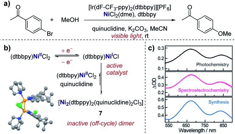
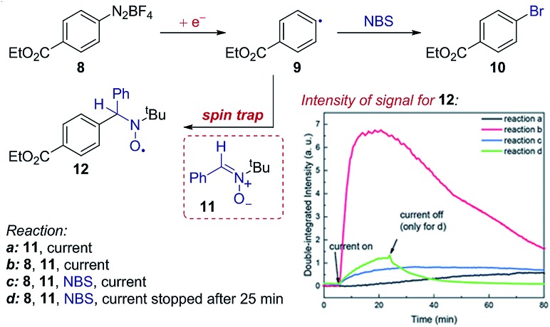
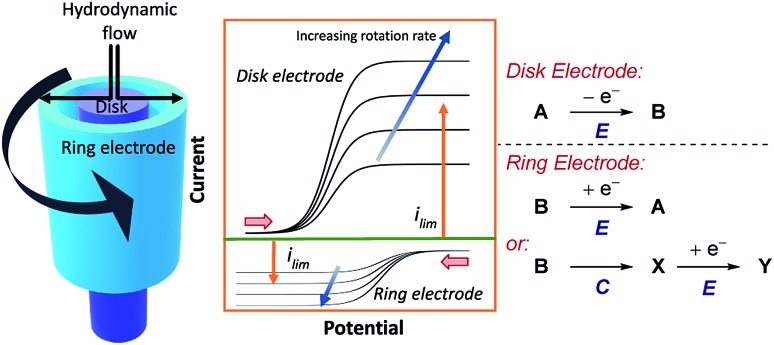
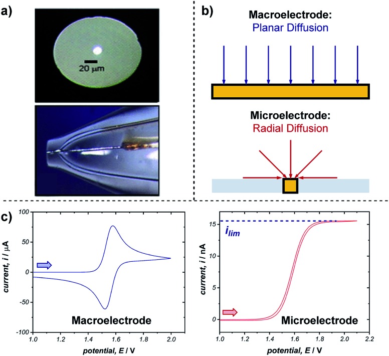
Monitoring reactive intermediates can provide vital information in the study of synthetic reaction mechanisms, enabling the design of new catalysts and methods. Many synthetic transformations are centred on the alteration of oxidation states, but these redox processes frequently pass through intermediates with short life-times, making their study challenging. A variety of electroanalytical tools can be utilised to investigate these redox-active intermediates: from voltammetry to in situ spectroelectrochemistry and scanning electrochemical microscopy. This perspective provides an overview of these tools, with examples of both electrochemically-initiated processes and monitoring redox-active intermediates formed chemically in solution. The article is designed to introduce synthetic organic and organometallic chemists to electroanalytical techniques and their use in probing key mechanistic questions.


Figures

Fig. 1
Fig. 1. (a) Example of a triangular potential waveform applied in the generation of a cyclic voltammogram. Representative CV responses for (b) a chemically reversible electron transfer (E mechanism), and (c) a chemically irreversible electron transfer in which the redox event is followed by a chemical reaction (EC mechanism). Two lines in (c) represent different rates of the following chemical reaction, where the dashed grey line has a rate constant of 0.1 s–1 and the solid red line a rate constant of 10 s–1 (with Ep/2 labelled for the latter). Arrows in figures demonstrate the direction of the scan, positive current represents oxidation.
Fig. 2
Fig. 2. (a) An example of CV used to monitor the speciation of the active titanocene catalyst (Cp2TiBr), depending on the equivalents of thiourea 3 added, utilised in a radical epoxide arylation by Gansäuer and co-workers. (b) Depiction of EC mechanism of titanocene catalyst. (c) CV responses with varying concentration of thiourea 3. Red arrow in figure demonstrates the direction of the scan, positive current represents oxidation. (c) Adapted with permission from ref. 12a. Copyright 2018 John Wiley and Sons.
Fig. 3
Fig. 3. CV responses displaying the dependence of the separation in potential of the two electron oxidation processes on the structure of the anilide, reported by Waldvogel and co-workers. (a) Depiction of the ECE mechanism of anilide oxidation. (b) CV responses of anilides 4 and 5. Red arrows in figures demonstrate the direction of the scan, positive current represents oxidation. (b) Adapted with permission from ref. 13. Copyright 2017 American Chemical Society.
Fig. 4
Fig. 4. Effect of the binding of Nd(OTf)3 to a ketone on the redox potential facilitating photoredox catalysed reduction, reported by Zeitler and co-workers. Blue arrow in figure demonstrates the direction of the scan, positive current represents oxidation. Adapted with permission from ref. 14. Copyright 2018 The Royal Society of Chemistry.
Fig. 5
Fig. 5. Nernstian dependence of the potential of the TEMPO redox couple on the concentration of azide demonstrates a 1 : 1 stoichiometry charge-transfer complex, reported by Lin and co-workers. Red arrow in figure demonstrates the direction of the scan, positive current represents oxidation. Adapted with permission from ref. 15. Copyright 2018 American Chemical Society.
Scheme 1
Scheme 1. Proposed anodic cyclisation of an amine with a dithioketene acetal, reported by Xu and Moeller [ref. 16a].
Fig. 6
Fig. 6. Representative CV responses for a catalytic (EC′) mechanism, demonstrating the change between peak-shaped response and plateau response as the substrate concentration increases. Simulations run with 0 equivalents (dashed red line), 10 equivalents (solid blue line) and 100 equivalents (solid black line) of substrate respectively. Arrow in figure demonstrates the direction of the scan, positive current represents oxidation.
Fig. 7
Fig. 7. (a) Evidence of electrocatalysis (EC′ mechanism) by a ligated Co(ii) species through oxidative addition of benzyl bromide, reported by the groups of Minteer and Sigman. Blue arrow in figure demonstrates the direction of the scan, positive current represents reduction. Adapted with permission from ref. 20. Copyright 2019 American Chemical Society. (b) Determination of catalytic Ru oxidation states from the increase in current due to catalysis, reported by the groups of Du Bois, Waymouth, Sigman and Zare. Blue arrow in figure demonstrates the direction of the scan, positive current represents reduction. Adapted with permission from ref. 21. Copyright 2019 American Chemical Society.
Fig. 8
Fig. 8. (a) Kinetic measurements of the Co(i) disproportionation rate constant from CV studies, reported by the groups of Minteer and Sigman. (b) Variable scan rate CV responses demonstrating the changing ipa/ipc ratio due to chemical depletion of Co(i) in the EC mechanism. Blue arrow in figure demonstrates the direction of the scan, positive current represents reduction. (c) Second order rate plot used to determine the rate constant. (b and c) Adapted with permission from ref. 20. Copyright 2019 American Chemical Society.
Fig. 9
Fig. 9. (a–c) Kinetic studies of a cooperative electrocatalytic alcohol oxidation by measuring the catalytic current on a CV response as a function of substrate/catalyst concentration, reported by Badalyan and Stahl. (d) Proposed catalytic cycle for alcohol oxidation. Blue arrow in figure demonstrates the direction of the scan, positive current represents oxidation. (a–c) Adapted with permission from ref. 28. Copyright 2016 Springer Nature.
Fig. 10
Fig. 10. (a) Kinetic studies on the rate of oxidative addition of chloroacetonitrile 6 to cobalt(i) tetraphenylporphyrin (ligand depicted by the four nitrogen binding sites as circles), reported by Costentin et al. (b) The shift in the peak reduction potential by the concentration of chloroacetonitrile (RX) allowed determination of the rate constant. Red arrow in figure demonstrates the direction of the scan, positive current represents reduction. (b) Adapted with permission from ref. 35. Copyright 2013 The Royal Society of Chemistry.
Fig. 11
Fig. 11. (a) Staircase-shaped waveform of a SWV experiment. (b) “Bell-shaped” curve that results from the signal of a chemically reversible redox couple of an electroactive species in a SWV experiment (cf., representative CV response in Fig. 1b). Red arrow in (b) demonstrates the direction of the scan, positive current represents oxidation.
Fig. 12
Fig. 12. (a) SWV study of the electrochemical Birch reduction of naphthalene provides evidence for two sequential electron-transfer steps, consistent with an ECEC mechanism, reported by the groups of Baran, Minteer, Anderson and Neurock. (b) SWV response at two different pulse frequencies. Red arrow in figure demonstrates the direction of the scan, positive current represents reduction. (b) Adapted with permission from ref. 41. Copyright 2019 The American Association for the Advancement of Science.
Fig. 13
Fig. 13. (a) The conversion of the fluorination of 2,4-dinitrochlorobenzene (2,4-DNCB) can be monitored during the reaction by peak fitting reduction peaks using SWV, reported by Compton and co-workers. (b) The signal for 2,4-DNFB (dashes) can be obtained by subtracting the signal of 2,4-DNCB (solid) from the combined SWV signal (dots). Red arrow in figure demonstrates the direction of the scan, positive current represents oxidation. (b) Adapted with permission from ref. 42. Copyright 2002 John Wiley and Sons.
Fig. 14
Fig. 14. (a) Evidence for the formation of a mixed-valent Ni(i/ii) dimer (7) in the photochemical etherification of arylhalides, reported by Nocera and co-workers. (b) 7 was synthesised independently, and the corresponding UV-Vis profile found to match measurements made of the reaction mixture in both photochemical and spectroelectrochemical settings (c). (b and c) Adapted with permission from ref. 48. Copyright 2019 American Chemical Society.
Fig. 15
Fig. 15. EPR spectroelectrochemistry provides evidence for the formation of radical 9 following electrochemical reduction, and the subsequent reaction with N-bromosuccinimide (NBS), reported by Mo and co-workers. The formation of radical 9 is measured by the EPR signal of the long-lived radical 12 afforded by spin-trapping. Adapted with permission from ref. 50. Copyright 2018 The Royal Society of Chemistry.
Fig. 16
Fig. 16. RDE experiments provide evidence for a palladium-hydroxyl species as a catalytic intermediate in the Suzuki cross-coupling reaction, reported by Amatore et al. For inset in (b), x is the fractional conversion to Pd(0). (a) Adapted with permission from ref. 58. Copyright 2013 John Wiley and Sons. (b and c) Adapted with permission from ref. 57. Copyright 2011 John Wiley and Sons.
Fig. 17
Fig. 17. Schematic for an RRDE, demonstrating the typical current response at the disk electrode for a hypothetical oxidation reaction, followed by subsequent reduction at the ring. Each set of curves is separately measured by holding the potential at one electrode constant, whilst sweeping the potential at the other electrode across a given range. The figure displayed is thereby a composite of these two sets of experiments. Red arrows in figure demonstrate the direction of the scans, positive current represents oxidation.
Fig. 18
Fig. 18. (a) Optical microscope images of the top and side of an exemplar microelectrode. (b) The difference in transport to a macro- and microelectrode results in differing voltammetric responses, shown for a representative reversible oxidation in (c). Arrows in (c) demonstrate the direction of the scan, positive current represents oxidation. (a) Adapted with permission from ref. 65c. Copyright 2006 Institute of Physics Publishing.
Fig. 19
Fig. 19. (a) Measurement of oxidative addition rates of (Ph3P)4Pd(0), 17, into para-substituted aryliodides in toluene, reported by Amatore and Pflüger. (b) Voltammograms measuring the oxidation of Pd(0) with varying concentrations of 17 at a gold-disk microelectrode. (c) Calculation of the rate constant of oxidative addition for p-iodotoluene by plotting the concentration of 17 against time. (d) Hammett plot of rate constants with different aryliodides. Red arrow in (b) demonstrates the direction of the scan, positive current represents reduction. (b–d) Adapted with permission from ref. 67. Copyright 1990 American Chemical Society.
Fig. 20
Fig. 20. (a) Determination of both the diffusion coefficient and number of electrons transferred in the reduction of 19, by the groups of Baran, Minteer, Neurock and White. The current response upon application of a potential step is measured (b), and the slope of the plot in (c) allows determination of the diffusion coefficient. Positive current represents reduction. (b and c) Adapted with permission from ref. 39c. Copyright 2019 American Chemical Society.
Fig. 21
Fig. 21. Representative microelectrode voltammograms of the reduction of species A with unknown charge at a microelectrode, (a) in the presence of supporting electrolyte, and (b) in the absence of supporting electrolyte, wherein the limiting current of the voltammogram changes depending on the charge of the species undergoing reduction. (c) Measurement of the limiting current of the two-electron reduction of proposed complex 19 (cf., Fig. 20) with and without electrolyte demonstrates that the complex bears a positive charge, by White and co-workers [ref. 72]. Red arrows in figure demonstrate the direction of the scan, positive current represents reduction.
Fig. 22
Fig. 22. (a) Schematic of an SECM set-up, where the distance (d) between the substrate and tip electrodes can be changed to measure concentrations in solution. The concentration profile of species in the mediated reduction of iodobenzene (b), measured by Amatore et al. using SECM. (c) Schematic of concentration profiles. (d) Concentration profile of 22 with varying equivalents of 23, measured by changing the distance (d) between the two electrodes. (e) Comparative concentration profile of various species in solution. (c–e) Adapted with permission from ref. 75. Copyright 2001 John Wiley and Sons.
Fig. 23
Fig. 23. SECM study of the reduction of carbon dioxide allows detection of the short-lived radical anion intermediate, reported by Bard and co-workers. Blue arrow in figure demonstrates the direction of the scan, positive current represents reduction. Adapted with permission from ref. 78a. Copyright 2017 American Chemical Society.
Fig. 24
Fig. 24. (a) Schematic of a bipolar electrochemical set-up, demonstrating the formation of anodic and cathodic poles on a bipolar electrode (BPE). (b) Schematic of a split-bipolar electrode in a U-type cell. (c) Fluorination of triphenylmethane can occur at low concentrations of electrolyte solution utilising a BPE set-up, reported by Inagi and co-workers [ref. 83; poly(ethylene glycol) (PEG) additive required to solubilise CsF in acetonitrile].

References

    1. Yan M., Lo J. C., Edwards J. T., Baran P. S. J. Am. Chem. Soc. 2016;138:12692–12714. - PMC - PubMed
    1. Shaw M. H., Twilton J., MacMillan D. W. C. J. Org. Chem. 2016;81:6898–6926. - PMC - PubMed
    2. Skubi K. L., Blum T. R., Yoon T. P. Chem. Rev. 2016;116:10035–10074. - PMC - PubMed
    3. Romero N. A., Nicewicz D. A. Chem. Rev. 2016;116:10075–10166. - PubMed
    4. Staveness D., Bosque I., Stephenson C. R. J. Acc. Chem. Res. 2016;49:2295–2306. - PMC - PubMed
    1. Yan M., Kawamata Y., Baran P. S. Chem. Rev. 2017;117:13230–13319. - PMC - PubMed
    2. Möhle S., Zirbes M., Rodrigo E., Gieshoff T., Wiebe A., Waldvogel S. R. Angew. Chem., Int. Ed. 2018;57:6018–6041. - PMC - PubMed
    3. Kärkäs M. D. Chem. Soc. Rev. 2018;47:5786–5865. - PubMed
    1. Bard A. J. and Faulkner L. R., Electrochemical Methods, John Wiley & Sons, Inc., Hoboken NJ, 2nd edn, 2001.
    2. Organic Electrochemistry, ed. O. Hammerich and B. Speiser, Taylor & Francis Group, Boca Raton FL, 5th edn, 2016.
    3. Jutand A. Chem. Rev. 2008;108:2300–2347. - PubMed
    4. Savéant J.-M. Chem. Rev. 2008;108:2348–2378. - PubMed
    5. Costentin C., Drouet S., Passard G., Robert M., Savéant J.-M. J. Am. Chem. Soc. 2013;135:9023–9031. - PubMed
    6. Rountree E. S., McCarthy B. D., Eisenhart T. T., Dempsey J. L. Inorg. Chem. 2014;53:9983–10002. - PubMed
    7. Rountree E. S., Martin D. J., McCarthy B. D., Dempsey J. L. ACS Catal. 2016;6:3326–3335.
    8. Elgrishi N., McCarthy B. D., Rountree E. S., Dempsey J. L. ACS Catal. 2016;6:3644–3659.
    9. Passard G., Ullman A. M., Brodsky C. N., Nocera D. G. J. Am. Chem. Soc. 2016;138:2925–2928. - PubMed
    10. Costentin C., Robert M., Savéant J.-M. Curr. Opin. Electrochem. 2017;2:26–31.
    1. Savéant J.-M., Elements of Molecular and Biomolecular Electrochemistry, John Wiley & Sons, Inc., Hoboken NJ, 2006.
    2. Elgrishi N., Rountree K. J., McCarthy B. D., Rountree E. S., Eisenhart T. T., Dempsey J. L. J. Chem. Educ. 2018;95:197–206.
    3. Graham D. J., Standard Operating Procedures for Cyclic Voltammetry, 2nd edn, 2018.