. 2019 Jan 14;7(2):198-209.
doi: 10.1039/C8TB01524D. Epub 2018 Nov 16.

Near-infrared fluorescent probes based on TBET and FRET rhodamine acceptors with different p K a values for sensitive ratiometric visualization of pH changes in live cells

Affiliations

Near-infrared fluorescent probes based on TBET and FRET rhodamine acceptors with different p K a values for sensitive ratiometric visualization of pH changes in live cells

Jianbo Wang et al. J Mater Chem B. .

Abstract

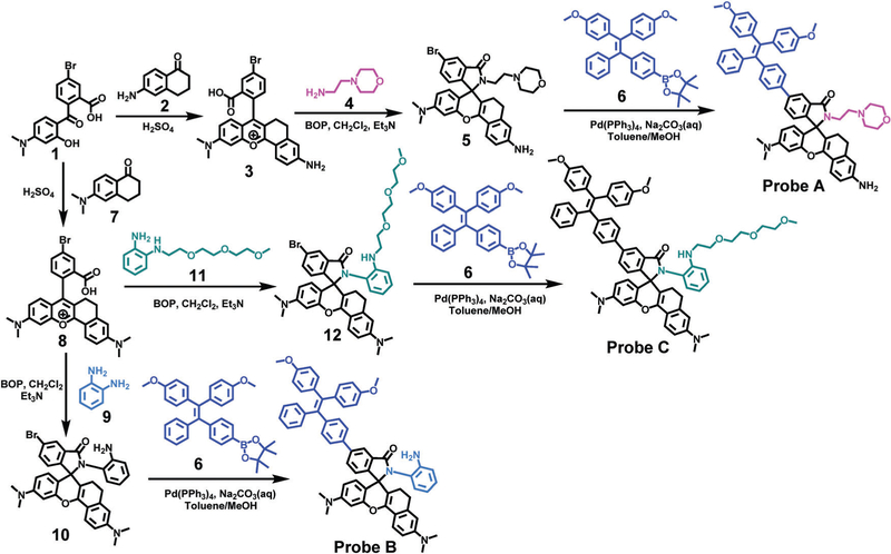
Three near-infrared ratiometric fluorescent probes (A-C) based on TBET and FRET near-infrared rhodamine acceptors with different pK a values were designed and synthesized to achieve sensitive ratiometric visualization of pH variations in lysosomes in visible and near-infrared channels. Tetraphenylethene (TPE) was bonded to near-infrared rhodamine dyes through short electrical π -conjugation linkers to prevent an aggregation-caused quenching (ACQ) effect and allow highly efficient energy transfer of up to 98.9% from TPE donors to rhodamine acceptors. Probes A-C respond to pH variation from 7.4 to 3.0 in both buffer solutions and live cells with significant decreases of donor fluorescence and concomitant extraordinary increases of rhodamine acceptor fluorescence because of highly efficient energy transfer. In addition, probe C is capable of determining pH fluctuations in live cells treated with chloroquine. The probes show good photostability, excellent cell membrane permeability, high selectivity to pH, and two well-resolved emission peaks to ensure accurately comparative and quantitative analyses of intracellular pH changes.


Conflict of interest statement

Conflicts of interest There are no conflicts to declare.

Figures

Fig. 1
Fig. 1
Absorption and fluorescence spectra of 10 µM probe A in 10 mM citrate buffers with the pH range from 7.58 to 2.38 containing 30% acetonitrile under TPE excitation at 405 nm. The ratio between the TPE donor fluorescence and the rhodamine acceptor fluorescence versus pH values for the probe.
Fig. 2
Fig. 2
Absorption and fluorescence spectra of 10 µM probe B in 10 mM citrate buffers with the pH range from 7.58 to 2.38 containing 30% acetonitrile under TPE excitation at 405 nm. The ratio between the TPE donor fluorescence and the rhodamine acceptor fluorescence versus pH values for the probe.
Fig. 3
Fig. 3
Absorption and fluorescence spectra of 10 µM probe C in 10 mM citrate buffers with the pH range from 7.58 to 2.38 containing 30% acetonitrile under TPE excitation at 405 nm. The ratio between the TPE donor fluorescence and the rhodamine acceptor fluorescence versus pH values for the probe.
Fig. 4
Fig. 4
Mercury drawing of the optimized geometry for probe A (left) and probe AH+ (right). H atoms are omitted for clarity. Grey: carbon; red: oxygen; blue: nitrogen. The labels apply to equivalent atom positions with the other probes which have different groups attached to the rhodamine moiety.
Fig. 5
Fig. 5
Difference density illustrations as isosurfaces of probes for the excited states indicated. Blue/red areas indicate values for different densities of ±1.00e−5 for the middle column and ±5.00e−5 for the outer ones, see the scale on top of the illustration.
Fig. 6
Fig. 6
Cellular fluorescence images of 15 µM probe C incubated with HeLa cells in 10 mM citrate buffers with the pH range from 3.5 to 7.0 in the presence of 5 µg mL nigericin. The blue channel in the first row was collected from 475 to 525 nm, and two NIR channels (pseudo-colored as red and green for clarity) in the second and third rows were collected from 650 to 700 nm under excitation of the TPE donor and the rhodamine acceptor at 405 nm and 559 nm, respectively. A confocal fluorescence microscope was employed to obtain fluorescence images at 60× magnification with scale bars of 50 µM.
Fig. 7
Fig. 7
Cellular fluorescence intensities of TPE donors and rhodamine acceptors in probes A (left), B (middle) and C (right) in 10 mM citrate buffers with the pH range from 3.5 to 7.0 having 5 µg mL nigericin under TPE excitation at 405 nm. Statistical analysis of the confocal imaging data in Fig. 6 and Fig. S45, S47 (ESI†) generates the donor and acceptor fluorescence intensities of probes A–C in HeLa live cells.
Fig. 8
Fig. 8
Cellular fluorescence images of 20 µM probe C incubated with HeLa cells in 10 mM citrate buffers with pH 7.4 in the absence and in the presence of 100 and 200 µM chloroquine. The blue channel in the first column was obtained from 475 to 525 nm, and two NIR channels (pseudo-colored as red and green for clarity) in the second and third columns were got from 650 to 700 nm under excitation of the TPE donor and the rhodamine acceptor at 405 nm and 559 nm, respectively. A confocal fluorescence microscope was used to acquire the images at 60× magnification with scale bars of 50 µM.
Scheme 1
Scheme 1
Chemical structure responses of fluorescent probes to pH changes with p-conjugation alternations.
Scheme 2
Scheme 2
Syntheses of probes A–C.

References

    1. Kaur B, Kaur N and Kumar S, Coord. Chem. Rev, 2018, 358, 13–69.
    1. Sivaraman G, Iniya M, Anand T, Kotla NG, Sunnapu O, Singaravadivel S, Gulyani A and Chellappa D, Coord. Chem. Rev, 2018, 357, 50–104.
    1. Gupta A and Kumar N, RSC Adv, 2016, 6, 106413–106434.
    1. Zhang RQ, Yan FY, Huang YC, Kong DP, Ye QH, Xu JX and Chen L, RSC Adv, 2016, 6, 50732–50760.
    1. Zhu H, Fan JL, Wang BH and Peng XJ, Chem. Soc. Rev, 2015, 44, 4337–4366. - PubMed

Publication types