

Hepatic iron overload identified by magnetic resonance imaging-based T2* is a predictor of non-diagnostic elastography
- PMID: 31367546
- PMCID: PMC6629566
- DOI: 10.21037/qims.2019.05.13
Hepatic iron overload identified by magnetic resonance imaging-based T2* is a predictor of non-diagnostic elastography
Abstract
Background: Magnetic resonance elastography (MRE) is a non-invasive test used to assess liver stiffness and fibrosis in chronic liver disease, which includes systemic iron overload. However, iron deposition by itself is associated with technical failure of MRE of the liver which necessitates the use of invasive liver biopsy as an alternative monitoring method for these patients. T2*-weighted magnetic resonance imaging (T2*) is a reliable modality to asses for hepatic as well as total body iron overload. Therefore, we aimed to determine a cutoff value on the T2* reading at which MRE would no longer provide accurate stiffness measurements in patients with iron overload.
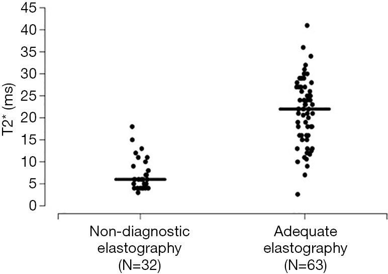
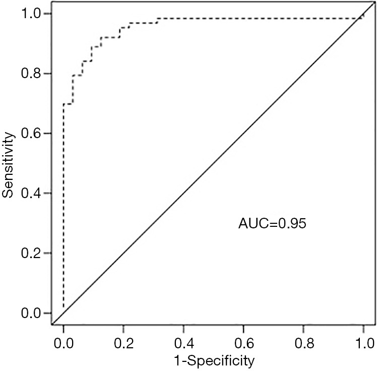
Methods: Ninety-five patients with iron overload who underwent MRE at our institution, between 2010 and 2017 were reviewed retrospectively. We compared T2* values between patients with adequate elastography (N=63) versus those with non-diagnostic elastography (N=32). We additionally examined the ability of T2* to predict the likelihood of non-diagnostic elastography by estimating area under the ROC curve (AUC).
Results: T2* was significantly different between patients with and without an adequate elastography (P<0.0001) and predicted occurrence of non-diagnostic elastography with an AUC of 0.95. All patients with a non-diagnostic elastography had a T2* value below 20 milliseconds (ms), and correspondingly 55% of the patients with a T2* value below 20 ms had a non-diagnostic elastography. The subgroups of patients with a T2* value ≤10, ≤8, and ≤6 ms, had a higher likelihood of non-diagnostic elastography (87%, 92%, and 95%, respectively).
Conclusions: T2* can be used to accurately predict which patients are most likely to have a non-diagnostic elastography reading. T2* of 20 ms or lower reflects a higher likelihood of non-diagnostic elastography.
Keywords: Hemochromatosis; T2* cut-off value; magnetic resonance elastography (MRE).
Conflict of interest statement
Conflicts of Interest: The authors have no conflicts of interest to declare.
Figures
References
LinkOut - more resources
Full Text Sources