. 2019 Jun;9(6):1087-1094.
doi: 10.21037/qims.2019.06.09.

A comparison of ultra-high-resolution CT target scan versus conventional CT target reconstruction in the evaluation of ground-glass-nodule-like lung adenocarcinoma

Affiliations

A comparison of ultra-high-resolution CT target scan versus conventional CT target reconstruction in the evaluation of ground-glass-nodule-like lung adenocarcinoma

Yanyan Zhu et al. Quant Imaging Med Surg. 2019 Jun.

Abstract

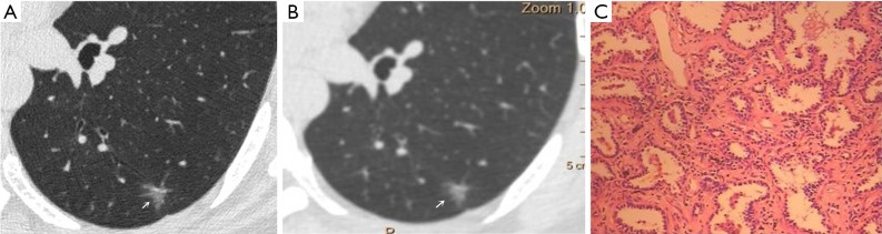
Background: The aim of this study was to determine whether the clinical value of scanned computed tomography (CT) images is higher when using ultra-high-resolution CT (U-HRCT) target scanning than conventional CT target reconstruction scanning in the evaluation of ground-glass-nodule (GGN)-like lung adenocarcinoma.

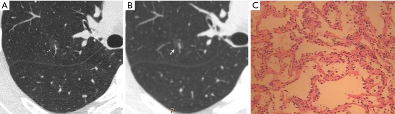
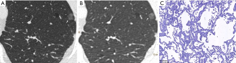
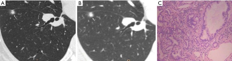
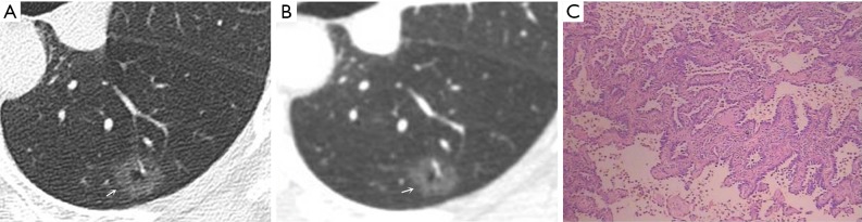
Methods: A total of 91 consecutive patients with isolated GGN-like lung adenocarcinoma were included in this study from April 2017 to June 2018. U-HRCT and conventional CT scans were conducted in all enrolled patients. Two experienced thoracic radiologists independently assessed image quality and made diagnoses. Based on the pathological results, the accuracies of U-HRCT target scanning and conventional CT target reconstruction for detecting morphological features on CT, including spiculation of GGNs, bronchial vascular bundles, solid components in the nodules, burr, vacuole, air bronchial signs, and fissure distortion, were calculated. All statistical analyses were performed using SPSS 17.0 software. Enumeration data were tested using the Chi-square test. A P value of <0.05 was considered statistically significant.

Results: When both techniques were compared with the pathological findings, the detection rate for CT images obtained using U-HRCT target scanning and conventional CT target reconstruction with regard to the spiculation of GGNs, bronchial vascular bundles, and solid components in the nodules were 78% vs. 61.5%, 72.5% vs. 54.9%, 65.9% vs. 49.5%, respectively. The presence of the spiculation of GGNs, bronchial vascular bundles, and solid components in the nodules in U-HRCT target scanning was significantly higher than that in conventional CT target reconstruction (all P<0.05). However, no significant difference was observed between the two techniques with regard to the burr, vacuole, air bronchial signs, and fissure distortion (all P>0.05).

Conclusions: When viewing GGNs, the detection rate was higher for U-HRCT target scanning than for conventional CT target reconstruction, and this improvement significantly enhanced the diagnostic accuracy of early lung adenocarcinoma.

Keywords: Ultra-high-resolution CT target scan (U-HRCT target scan); conventional CT target reconstruction; ground-glass opacity; lung adenocarcinoma.


Conflict of interest statement

Conflicts of Interest: The authors have no conflicts of interest to declare.

Figures

Figure 1
Figure 1
Male, 53 years old. A U-HRCT target scan image shows clear speculation and a bronchial vascular bundle in the ground-glass nodule (A). A conventional CT target reconstruction image shows unclear spiculation and bronchial vascular bundles in the ground-glass nodule (B). The pathological image depicts the tumour cells being sparsely arranged along the alveolar wall, thus presenting a ground-glass nodule and a clearer tumour-lung interface on CT imaging. The pathological results confirmed the presence of atypical adenomatous hyperplasia (C, HE staining, 200×). U-HRCT, ultra-high-resolution computed tomography. The arrows point out the lesions.
Figure 2
Figure 2
Male, 65 years old. A U-HRCT target scan image (A) and a conventional CT target reconstruction image (B) show clear and sharp spiculation of the ground-glass nodule. The pathological image indicates that the tumour cells are tightly arranged along the alveolar wall, thus presenting a ground-glass nodule and a clearer tumour-lung interface on CT imaging. The pathological results confirmed the presence of adenocarcinoma in situ (C, HE staining, 100×). U-HRCT, ultra-high-resolution computed tomography. The arrows point out the lesions.
Figure 3
Figure 3
Female, 48 years old. U-HRCT target scan (A) shows the clear spiculation of the ground-glass nodule. The central solid component in the nodule is clearly separated from the surrounding ground-glass opacity, and the adjacent fissure is distorted and thickened. A conventional CT target reconstruction (B) shows that the central solid component in the nodule is unclearly separated from the surrounding ground-glass opacity, while the spiculation of the ground-glass nodule is blurred. The adjacent fissure is distorted, and the thickness is not obvious. The pathological image indicates tumour infiltration into the matrix leading to the contraction of the elastic tissues of the lung with the adjacent fissure distortion, thus presenting a central solid component in the nodule on CT imaging. The pathological results confirmed the presence of microinvasive adenocarcinoma (C, HE staining, 100×). U-HRCT, ultra-high-resolution computed tomography. The arrows point out the lesions.
Figure 4
Figure 4
Female, 57 years old. A U-HRCT target scan (A) shows a vacuole sign in the GGN. A conventional CT target reconstruction (B) shows that the vacuole sign in the nodule is unclearly separated from the surrounding ground-glass opacity. The pathological image indicates tumour infiltration into the matrix leading to the contraction of the elastic tissues of the lung, thus presenting a vacuole sign (normal lung tissue inside the nodule) on CT imaging. The pathological results confirmed the presence of invasive adenocarcinoma (C, HE staining, 200×). U-HRCT, ultra-high-resolution computed tomography; GGN, ground-glass nodule. The arrows point out the lesions.
Figure 5
Figure 5
Female, 49 years old. A U-HRCT target scan (A) clearly shows the bronchial vascular bundle inside the nodule and burr sign at the nodule edge. A conventional CT target reconstruction (B) shows the uneven density of the nodule, but the bronchial vascular bundle and burr sign are unclear. The pathological image indicates tumour infiltration into the matrix leading to the contraction of the elastic tissues of the lung, with the contracted fibres pulling the surrounding normal lung tissue and the bronchial vascular bundle, thus presenting a burr sign and a bronchial vascular bundle on CT imaging. The pathological results confirmed the presence of invasive adenocarcinoma (C, HE staining, 200×). U-HRCT, ultra-high-resolution computed tomography. The arrows point out the lesions.

Similar articles

Cited by

References

    1. Travis WD, Brambilla E, Riely GJ. New pathologic classification of lung cancer: relevance for clinical practice and clinical trials. J Clin Oncol 2013;31:992-1001. 10.1200/JCO.2012.46.9270 - DOI - PubMed
    1. Todo G, Ito H, Nakano Y, Dodo Y, Maeda H, Murata K, Odori T, Torizuka K, Izumi T, Oshima S. High resolution CT (HR-CT) for the evaluation of pulmonary peripheral disorders. Rinsho Hoshasen 1982;27:1319-26. - PubMed
    1. Carter D, Vazquez M, Flieder DB, Brambilla E, Gazdar A, Noguchi M, Travis WD, Kramer A, Yip R, Yankelevitz DF, Henschke CI. Comparison of pathologic findings of baseline and annual repeat cancers diagnosed on CT screening. Lung Cancer 2007;56:193-9. 10.1016/j.lungcan.2006.12.001 - DOI - PubMed
    1. Goo JM, Park CM, Lee HJ. Ground-glass nodules on chest CT as imaging biomarkers in the management of lung adenocarcinoma. AJR Am J Roentgenol 2011;196:533-43. 10.2214/AJR.10.5813 - DOI - PubMed
    1. Henschke CI, Yankelevitz DF, Mirtcheva R, McGuinness G, McCauley D, Miettinen OS. CT screening for lung cancer: frequency and significance of part-solid and nonsolid nodules. AJR Am J Roentgenol 2002;178:1053-7. 10.2214/ajr.178.5.1781053 - DOI - PubMed