

Short-term changes in muscle activity and jaw movement patterns after orthognathic surgery in skeletal Class III patients with facial asymmetry
- PMID: 31367580
- PMCID: PMC6658898
- DOI: 10.4041/kjod.2019.49.4.254
Short-term changes in muscle activity and jaw movement patterns after orthognathic surgery in skeletal Class III patients with facial asymmetry
Abstract
Objective: To evaluate the short-term changes in masticatory muscle activity and mandibular movement patterns after orthognathic surgery in skeletal Class III patients with facial asymmetry.
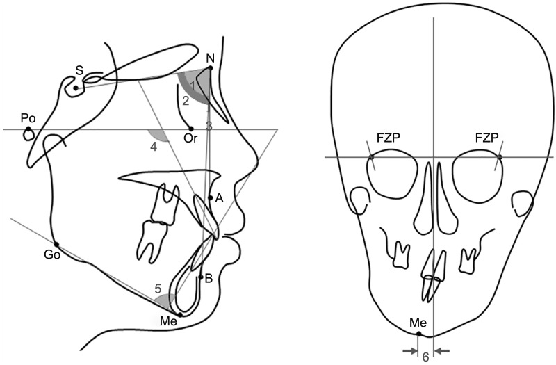
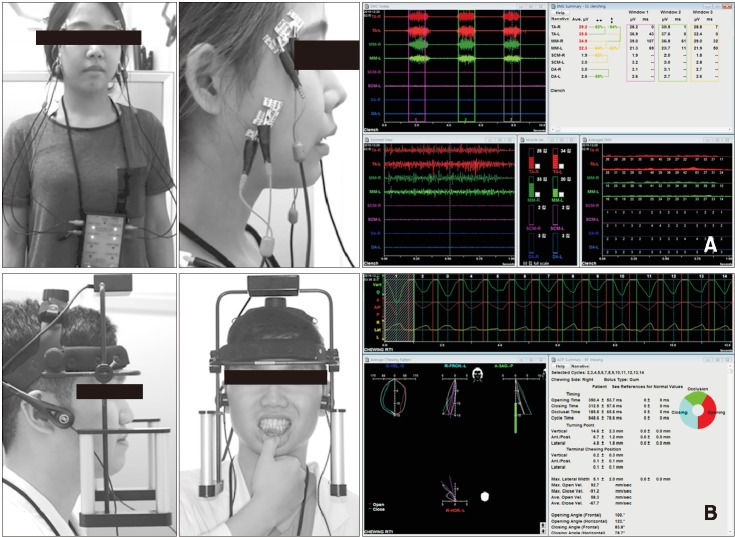
Methods: Twenty-seven skeletal Class III adult patients were divided into two groups based on the degree of facial asymmetry: the experimental group (n = 17 [11 male and 6 female]; menton deviation ≥ 4 mm) and control group (n = 10 [4 male and 6 female]; menton deviation < 1.6 mm). Cephalography, electromyography (EMG) for the anterior temporalis (TA) and masseter muscles (MM), and mandibular movement (range of motion [ROM] and average chewing pattern [ACP]) were evaluated before (T0) and 7 to 8 months (T1) after the surgery.
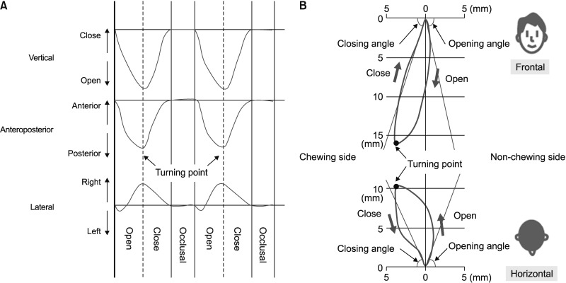
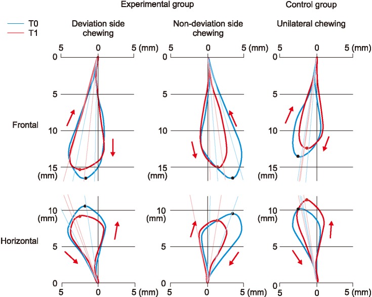
Results: There were no significant postoperative changes in the EMG potentials of the TA and MM in both groups, except in the anterior cotton roll biting test, in which the masticatory muscle activity had changed into an MM-dominant pattern postoperatively in both groups. In the experimental group, the amount of maximum opening, protrusion, and lateral excursion to the non-deviated side were significantly decreased. The turning point tended to be shorter and significantly moved medially during chewing in the non-deviated side in the experimental group.
Conclusions: In skeletal Class III patients with facial asymmetry, the EMG activity characteristics recovered to presurgical levels within 7 to 8 months after the surgery. Correction of the asymmetry caused limitation in jaw movement in terms of both ROM and ACP on the non-deviated side.
Keywords: Class III orthognathic surgery; Electromyography; Facial asymmetry; Jaw movement.
Conflict of interest statement
CONFLICTS OF INTEREST: No potential conflict of interest relevant to this article was reported.
Figures
Similar articles
-
Occlusal function and electromyographic activity of masticatory muscles in skeletal Class III patients with different patterns of mandibular asymmetry.J Oral Rehabil. 2023 Apr;50(4):276-285. doi: 10.1111/joor.13412. Epub 2023 Jan 19. J Oral Rehabil. 2023. PMID: 36597189
-
Influence of surgical orthodontic treatment on masticatory function in skeletal Class III patients.J Oral Rehabil. 2015 Oct;42(10):733-41. doi: 10.1111/joor.12307. Epub 2015 May 15. J Oral Rehabil. 2015. PMID: 25975774
-
Alteration of masticatory electromyographic activity and stability of orthognathic surgery in patients with skeletal class III malocclusion.J Oral Maxillofac Surg. 2013 Jul;71(7):1249-60. doi: 10.1016/j.joms.2013.01.002. Epub 2013 Apr 4. J Oral Maxillofac Surg. 2013. PMID: 23562358
-
Pharyngeal Airway Morphology in Skeletal Class III With Mandibular Asymmetry is Improved After Bimaxillary Orthognathic Surgery.J Oral Maxillofac Surg. 2021 May;79(5):1107-1121. doi: 10.1016/j.joms.2021.01.001. Epub 2021 Jan 9. J Oral Maxillofac Surg. 2021. PMID: 33549539
-
Changes in 3-Dimensional Measurements of Masseter Muscle After Orthognathic Surgery in Patients with Facial Asymmetry.Aesthetic Plast Surg. 2024 Oct;48(19):3751-3757. doi: 10.1007/s00266-024-04309-2. Epub 2024 Aug 26. Aesthetic Plast Surg. 2024. PMID: 39187590 Free PMC article.
Cited by
-
Computerized Assessment of Occlusion and Muscle Activity during Use of a Multilayer Clear Retainer: A Preliminary Study.Sensors (Basel). 2021 Jan 13;21(2):541. doi: 10.3390/s21020541. Sensors (Basel). 2021. PMID: 33451097 Free PMC article.
-
[Diagnostic and treatment alternatives for the correction of facial asymmetries: a literature review].Rev Cient Odontol (Lima). 2022 Mar 30;10(1):e098. doi: 10.21142/2523-2754-1001-2022-098. eCollection 2022 Jan-Mar. Rev Cient Odontol (Lima). 2022. PMID: 38389908 Free PMC article. Review. Spanish.
-
Comparison of the bite force and occlusal contact area of the deviated and non-deviated sides after intraoral vertical ramus osteotomy in skeletal Class III patients with mandibular asymmetry: Two-year follow-up.Korean J Orthod. 2022 May 4;52(3):172-81. doi: 10.4041/kjod21.236. Online ahead of print. Korean J Orthod. 2022. PMID: 35504730 Free PMC article.
-
Changes in the electromyographic activity of masticatory muscles in patients undergoing bimaxillary surgery.Acta Odontol Scand. 2025 Apr 22;84:182-190. doi: 10.2340/aos.v84.43408. Acta Odontol Scand. 2025. PMID: 40260992 Free PMC article.
References
-
- Castelo PM, Gavião MB, Pereira LJ, Bonjardim LR. Masticatory muscle thickness, bite force, and occlusal contacts in young children with unilateral posterior crossbite. Eur J Orthod. 2007;29:149–156. - PubMed
-
- Kiliaridis S, Mahboubi PH, Raadsheer MC, Katsaros C. Ultrasonographic thickness of the masseter muscle in growing individuals with unilateral crossbite. Angle Orthod. 2007;77:607–611. - PubMed
-
- Nie Q, Kanno Z, Xu T, Lin J, Soma K. Clinical study of frontal chewing patterns in various crossbite malocclusions. Am J Orthod Dentofacial Orthop. 2010;138:323–329. - PubMed
-
- Rilo B, da Silva JL, Mora MJ, Cadarso-Suárez C, Santana U. Unilateral posterior crossbite and mastication. Arch Oral Biol. 2007;52:474–478. - PubMed
-
- Hashimoto T, Kuroda S, E L, Tanimoto Y, Miyawaki S, Takano-Yamamoto T. Correlation between craniofacial and condylar path asymmetry. J Oral Maxillofac Surg. 2008;66:2020–2027. - PubMed
LinkOut - more resources
Full Text Sources