

Regression of Sebaceous Carcinoma of the Eyelid after a Small Incisional Biopsy: Report of Two Cases
- PMID: 31367586
- PMCID: PMC6615332
- DOI: 10.1159/000490706
Regression of Sebaceous Carcinoma of the Eyelid after a Small Incisional Biopsy: Report of Two Cases
Abstract
Purpose: To report 2 cases of regression of sebaceous carcinoma of the eyelid after a small incisional biopsy.
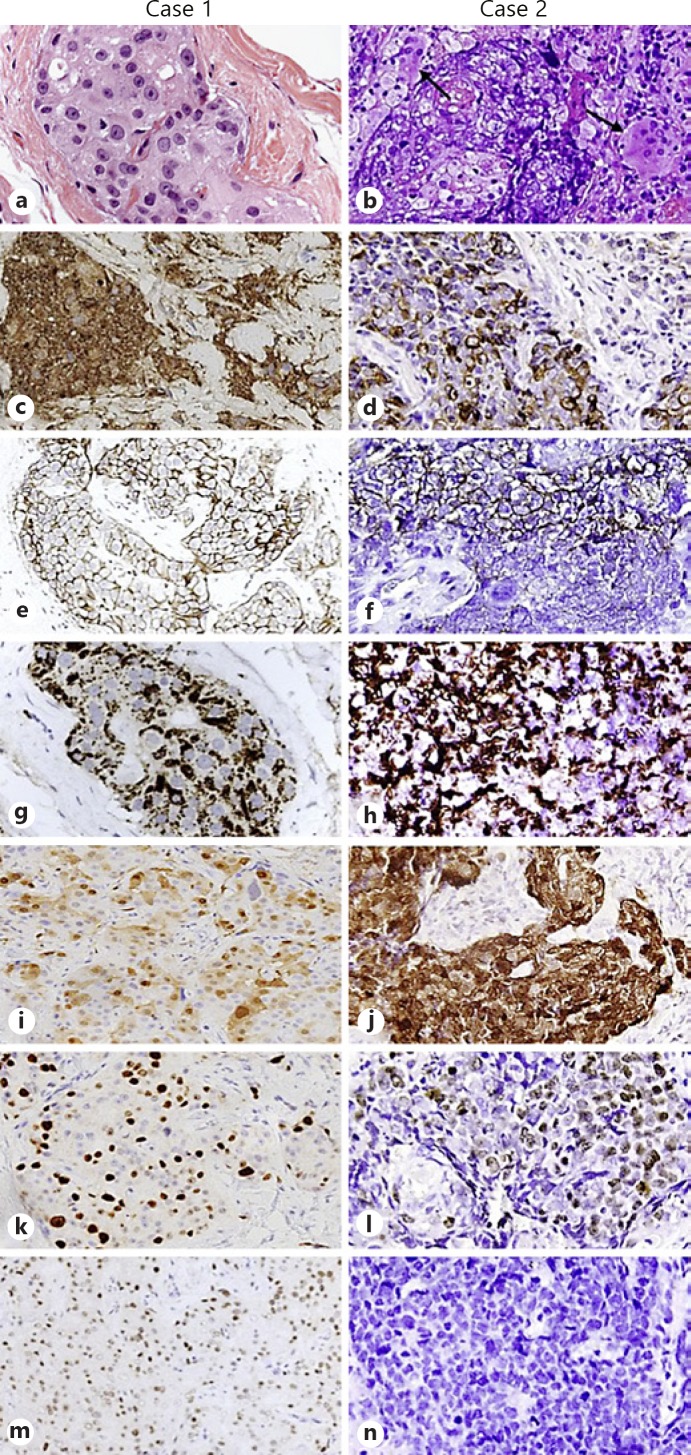
Methods: Clinical, imaging, and histopathological findings are presented, with a literature review on regressing ocular tumors.
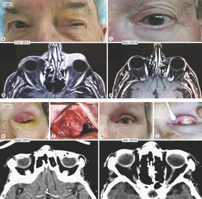
Results: Our first patient was a 79-year-old man who presented with a 10-month history of progressive left upper eyelid ptosis caused by an eyelid tumor with orbital involvement and confirmed on magnetic resonance imaging. Our second patient was a 70-year-old woman who presented with ptosis with a left upper eyelid mass. Both patients underwent a small incisional biopsy of their lesion. The histopathological diagnoses in both cases were consistent with sebaceous carcinoma. Both patients refused exenteration. Follow-up clinical examination and imaging disclosed total regression of the ptosis and of the neoplasm with no sign of recurrence in both patients over a 4-year period for Case 1 and a 7-year period for Case 2.
Conclusion: Regression following incisional biopsy of basal cell, squamous cell, and Merkel cell carcinoma, including of the eyelid, is well documented. To the best of our knowledge, our 2 cases of sebaceous carcinoma are the first to be reported with total involution clinically and on imaging of the tumor following partial incisional biopsy.
Keywords: Exenteration; Eyelid; Orbit; Ptosis; Sebaceous carcinoma; Small incisional biopsy; Spontaneous regression.
Conflict of interest statement
The authors have no financial conflict of interest to disclose. All of the authors have seen and given their approval for the submission of this paper. They warrant that this article is the authors' original work, has not received prior publication, and is not under consideration for publication elsewhere.
Figures
Similar articles
-
Selective removal of sebaceous gland carcinoma of the lower eyelid with orbital infiltration.Ophthalmic Surg Lasers Imaging. 2007 Sep-Oct;38(5):413-6. doi: 10.3928/15428877-20070901-12. Ophthalmic Surg Lasers Imaging. 2007. PMID: 17955850
-
Sebaceous cell carcinoma of the eyelid and the human immunodeficiency virus.Ophthalmic Plast Reconstr Surg. 2000 May;16(3):206-10. doi: 10.1097/00002341-200005000-00007. Ophthalmic Plast Reconstr Surg. 2000. PMID: 10826761
-
Primary sebaceous carcinoma of lacrimal gland: A case report and review of literature.World J Clin Cases. 2018 Dec 26;6(16):1194-1198. doi: 10.12998/wjcc.v6.i16.1194. World J Clin Cases. 2018. PMID: 30613681 Free PMC article.
-
Sebaceous carcinoma of the eyelids: personal experience with 60 cases.Ophthalmology. 2004 Dec;111(12):2151-7. doi: 10.1016/j.ophtha.2004.07.031. Ophthalmology. 2004. PMID: 15582067 Review.
-
Radiation therapy for local control of eyelid sebaceous cell carcinoma: report of two cases and review of the literature.Ophthalmic Plast Reconstr Surg. 2000 May;16(3):211-5. doi: 10.1097/00002341-200005000-00008. Ophthalmic Plast Reconstr Surg. 2000. PMID: 10826762 Review.
References
-
- Swetter SM, Boldrick JC, Pierre P, Wong P, Egbert BM. Effects of biopsy-induced wound healing on residual basal cell and squamous cell carcinomas: rate of tumor regression in excisional specimens. J Cutan Pathol. 2003 Feb;30((2)):139–46. - PubMed
-
- Missotten GS, de Wolff-Rouendaal D, de Keizer RJ. Merkel cell carcinoma of the eyelid review of the literature and report of patients with Merkel cell carcinoma showing spontaneous regression. Ophthalmology. 2008 Jan;115((1)):195–201. - PubMed
-
- Nathanson Spontaneous regression of malignant melanoma: a review of the literature on incidence, clinical features, and possible mechanisms. Natl Cancer Inst Monogr. 1976 Nov;44:67–76. - PubMed
-
- Claudy AL, Viac J, Schmitt D, Alario A, Thivolet J. Identification of mononuclear cells infiltrating basal cell carcinomas. Acta Derm Venereol. 1976;56((5)):361–5. - PubMed
Publication types
LinkOut - more resources
Full Text Sources
