

Melanoma-Associated Spongiform Scleropathy Adjacent to a Choroidal Nevus
- PMID: 31367587
- PMCID: PMC6615339
- DOI: 10.1159/000494326
Melanoma-Associated Spongiform Scleropathy Adjacent to a Choroidal Nevus
Abstract
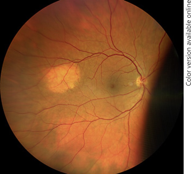
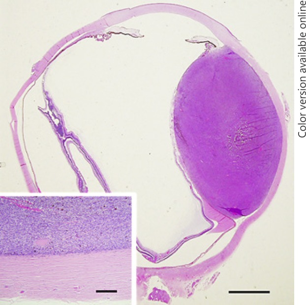
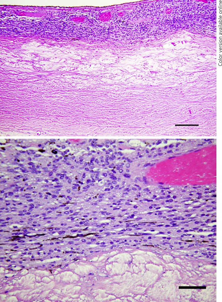
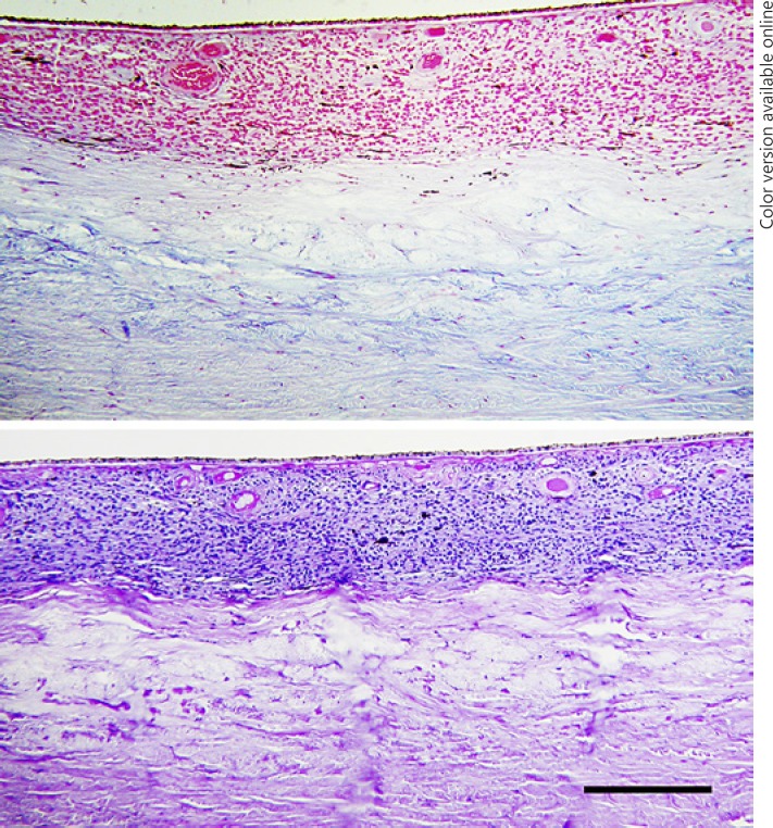
Melanoma-associated spongiform scleropathy contiguous to a choroidal nevus was an incidental finding in a 57-year-old woman whose eye was removed for a separate choroidal-ciliary body melanoma. All previously reported cases of melanoma-associated spongiform scleropathy, except for one, have been found adjacent to posterior uveal melanoma. The mechanism of scleral degeneration in melanoma-associated spongiform scleropathy is unknown. Few cases of posterior uveal nevi have been reported since the description of spongiform scleropathy, making assessment of the specificity of the degeneration to melanoma alone difficult. The presence of melanoma-associated scleropathy adjacent to a choroidal nevus indicates that the condition is not exclusively linked to posterior uveal melanoma.
Keywords: Choroidal nevus; Melanoma-associated spongiform scleropathy; Nevus; Scleral degeneration.
Conflict of interest statement
The authors have no conflicts of interest to declare.
Figures
References
-
- Alyahya GA, Heegaard S, Prause JU. Characterization of melanoma associated spongiform scleropathy. Acta Ophthalmol Scand. 2002 Jun;80((3)):322–6. - PubMed
-
- Alyahya GA. Melanoma associated spongiform scleropathy: characterization, biochemical and immunohistochemical studies. Acat Ophthalmol. 2008 Oct;86(Thesis 3):1–21. - PubMed
-
- Robert F, The Sclera . In: Eye Pathology. Heegaard S, Grosssnikluas HE, editors. Heidelberg: Springer; Springer. pp. pp. 155–72. Chapter 4.
-
- Alyahya GA, Ribel-Madsen SM, Heegaard S, Prause JU, Trier K. Melanoma-associated spongiform scleropathy: biochemical changes and possible relation to tumour extension. Acta Ophthalmol Scand. 2003 Dec;81((6)):625–9. - PubMed
Publication types
LinkOut - more resources
Full Text Sources
