

Case report of a sub-occluding thrombus in thoracic aorta: what is the origin?
- PMID: 31367736
- PMCID: PMC6764550
- DOI: 10.1093/ehjcr/ytz114
Case report of a sub-occluding thrombus in thoracic aorta: what is the origin?
Abstract
Background: Aortic thrombosis represents a consequence of atherosclerotic disease. In few cases, it can be secondary to large vessel or infective vasculitis. More rarely, aortic thrombosis is the manifestation of a primary malignant neoplasm of the aortic wall. Aortic angiosarcoma is a rare tumour, its clinical presentation is often non-specific and associated signs and symptoms may vary greatly. An early diagnosis is difficult to reach and the presence of metastatic disease is not uncommon at the time of diagnosis. The prognosis is poor overall.
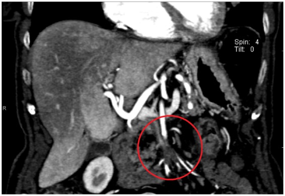
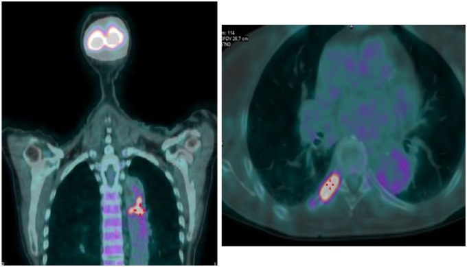
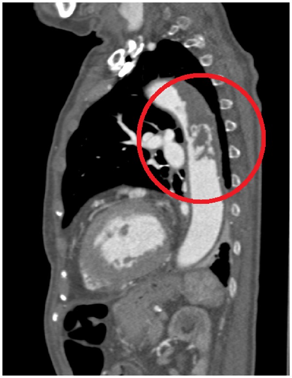
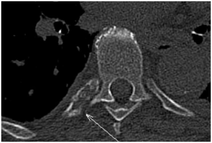
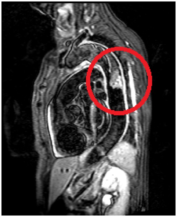
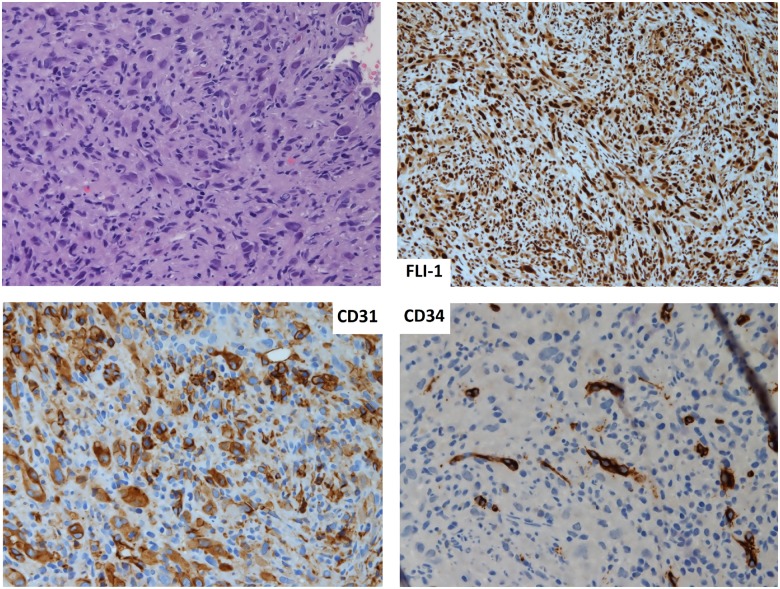
Case summary: We report the case of a female patient who presented to her GP because of fatigue, hyporexia, weight-loss, and anaemia. An ultrasound of the abdomen showed two small pancreatic lesions, confirmed and described as benign cystic pancreatic lesions on computed tomography (CT) imaging; an incidental thrombus in the superior mesenteric artery was also found on CT imaging. The thoracic CT identified a large thrombotic lesion in the descending thoracic aorta with significant narrowing of the aortic lumen and confirmed the presence of an osteolytic bone lesion on the VIII right rib, in the absence of atherosclerotic disease. Signs of increased metabolic activity in the aortic lumen and in the VIII posterior right rib were shown at a subsequent positron emission tomography. A CT-guided biopsy of the bone lesion was performed and at histology the diagnosis of metastatic angiosarcoma of the aortic wall was made.
Discussion: Aortic angiosarcoma is a rare cause of aortic thrombosis, to be taken into consideration in a patient with thrombotic lesions of the aorta in the absence of atherosclerotic disease. The differential diagnosis is difficult because of clinical presentation and radiological features similar to those of inflammatory aortic disease. In our case, the final diagnosis of angiosarcoma was made only by a biopsy of a bone metastatic lesion.
Keywords: Aortic angiosarcoma; Aortic thrombosis; Case report; Primary aortic tumour.
© The Author(s) 2019. Published by Oxford University Press on behalf of the European Society of Cardiology.
Figures

References
-
- Erbel R, Aboyans V, Boileau C, Bossone E, Bartolomeo RD, Eggebrecht H, Evangelista A, Falk V, Frank H, Gaemperli O, Grabenwöger M, Haverich A, Iung B, Manolis AJ, Meijboom F, Nienaber CA, Roffi M, Rousseau H, Sechtem U, Sirnes PA, Allmen RS, Vrints CJ; ESC Committee for Practice Guidelines. 2014 ESC Guidelines on the diagnosis and treatment of aortic diseases: document covering acute and chronic aortic diseases of the thoracic and abdominal aorta of the adult. The task force for the diagnosis and treatment of aortic diseases of the European Society of Cardiology (ESC). Eur Heart J 2014;35:2873–2926. - PubMed
-
- Brodowski, W. Primäres Sarkom der Aorta thoracica mit Verbreitung des Neugebildes in der unteren Körperhälfte. Jahresb Leistung Fortschr ges Med 1873;8:243–246.
-
- Rusthoven CG, Liu AK, Bui MM, Schefter TE, Elias AD, Lu X, Gonzalez RJ.. Sarcomas of the aorta: a systematic review and pooled analysis of published reports. Ann Vasc Surg 2014;28:515–525. - PubMed
-
- Thalheimer A, Fein M, Geissinger E, Franke S.. Intimal angiosarcoma of the aorta: report of a case and review of the literature. J Vasc Surg 2004;40:548–553. - PubMed
LinkOut - more resources
Full Text Sources