

A Novel ALK-THBS1 Fusion in a Laryngeal Inflammatory Myofibroblastic Tumour: A Case Report and Literature Review
- PMID: 31368077
- PMCID: PMC7235104
- DOI: 10.1007/s12105-019-01061-x
A Novel ALK-THBS1 Fusion in a Laryngeal Inflammatory Myofibroblastic Tumour: A Case Report and Literature Review
Abstract
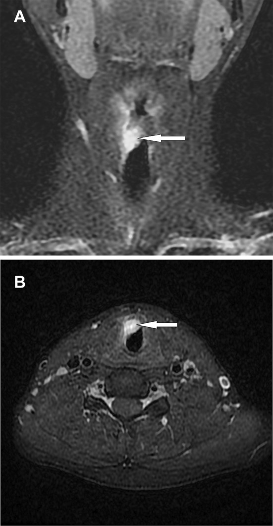
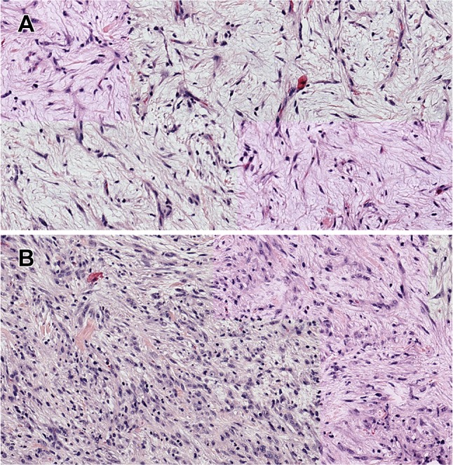
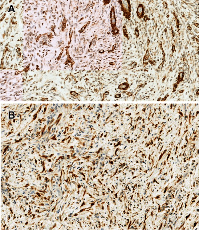
Inflammatory myofibroblastic tumor (IMT) is an uncommon neoplasm most frequently seen in the abdomino-pelvic region and lungs of children and young adults. Laryngeal tumors are rare. We present a case of a 23-year-old patient with a 5 month history of laryngitis and aphonia unresolved by corticotherapy. Laryngoscopy revealed a small, non-ulcerated, subepithelial, polypoid mass arising from the right vocal cord. The diagnosis of IMT with ALK expression was supported by histopathologic and molecular analysis. The THBS1 fusion partner was identified by RNA-sequencing analysis for the first time in a laryngeal IMT. This fusion partner has only been identified in six uterine IMTs thus far. Conservative excision of the lesion yielded excellent functional results for the patient. The voice was preserved and no recurrences were seen after 6 months of follow-up.
Keywords: ALK–THBS1 fusion; Anaplastic lymphoma kinase; Immunohistochemistry; Inflammatory myofibroblastic tumor; Laryngeal tumor.
Conflict of interest statement
The authors declare that there are no conflicts of interest regarding the publication of this paper.
Figures
References
-
- Coffin CM, Flechter JA. World Health Organization classification of tumors of soft tissue and bone. 4. Lyon: IARC; 2013. pp. 83–84.
-
- Ereño C, López JI, Grande J, Santaolalla F, Bilbao FJ. Inflammatory myofibroblastic tumour of the larynx. J Laryngol Otol. 2001;115:856–858. - PubMed
Publication types
MeSH terms
Substances
LinkOut - more resources
Full Text Sources
Miscellaneous
