. 2020 Apr;108(3):965-975.
doi: 10.1002/jbm.b.34449. Epub 2019 Jul 31.

System for application of controlled forces on dental implants in rat maxillae: Influence of the number of load cycles on bone healing

Affiliations

System for application of controlled forces on dental implants in rat maxillae: Influence of the number of load cycles on bone healing

Renan de Barros E Lima Bueno et al. J Biomed Mater Res B Appl Biomater. 2020 Apr.

Abstract

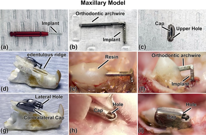
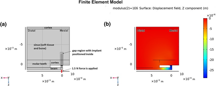
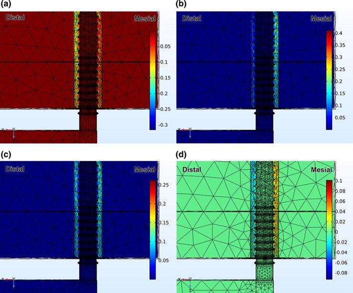
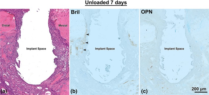
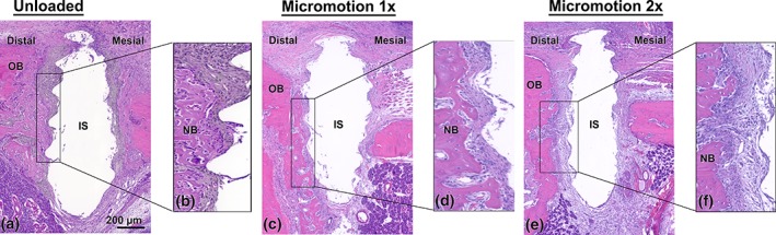
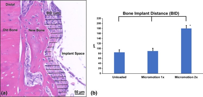
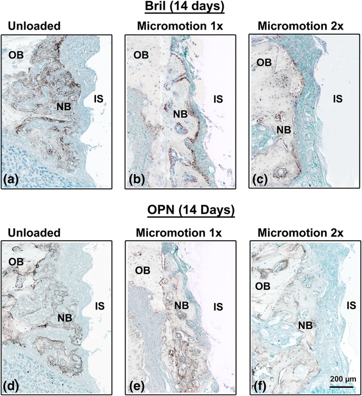
Experimental studies on the effect of micromotion on bone healing around implants are frequently conducted in long bones. In order to more closely reflect the anatomical and clinical environments around dental implants, and eventually be able to experimentally address load-management issues, we have developed a system that allows initial stabilization, protection from external forces, and controlled axial loading of implants. Screw-shaped implants were placed on the edentulous ridge in rat maxillae. Three loading regimens were applied to validate the system; case A no loading (unloaded implant) for 14 days, case B no loading in the first 7 days followed by 7 days of a single, daily loading session (60 cycles of an axial force of 1.5 N/cycle), and case C no loading in the first 7 days followed by 7 days of two such daily loading sessions. Finite element modeling of the peri-implant compressive and tensile strains plus histological and immunohistochemical analyses revealed that in case B any tissue damage resulting from the applied force (and related interfacial strains) did not per se disturb bone healing, however, in case C, the accumulation of damage resulting from the doubling of loading sessions severely disrupted the process. These proof-of-principle results validate the applicability of our system for controlled loading, and provide new evidence on the importance of the number of load cycles applied on healing of maxillary bone.

Keywords: bone; implant; loading; micromotion, maxilla, rat.


Conflict of interest statement

The authors have no conflicts of interest to report.

Figures

Figure 1
Figure 1
Micromotion system for rat maxillae; (a) photograph of the Retopin implant; (b) the implant welded to a titanium orthodontic arch wire; (c) the protective cap with a loading hole on its upper aspect; (d) implant system adapted and cemented on a dry maxilla and; (e, f) in situ on the edentulous ridge of the maxilla; (g) protective pediatric crown adapted to overhang the implant in a dry maxilla and (h, i) in situ. (c, h, and i) the loading hole can be positioned on the upper or (g) the lateral aspect of the cap. The holes in (g–i) were made bigger for illustration purposes
Figure 2
Figure 2
(a) Overall formulation of the FE model, showing idealized regions of molar teeth, cortical bone, sinus, titanium beam, gap, and implant. The thickness of the model is 1.5 mm into the page, with mesio‐distal and occluso‐apical dimensions as noted in the text. The mesial, distal, and superior surfaces of the model are fixed; (b) semi‐transparent view of a portion of the model showing z‐displacements after loading the end of the beam with 1.5 N, and with the modulus of the gap tissue equal to 1 MPa
Figure 3
Figure 3
(a) Distribution of 3rd principal strain (compressive) after loading the end of the beam with 1.5 N, and with the modulus of the gap tissue equal to 1 MPa; (b) distribution of 1st principal strain (tensile) after loading the end of the beam with 1.5 N, and with the modulus of the gap tissue equal to 1 MPa; (c) distribution of distortional strain after loading the end of the beam with 1.5 N, and with the modulus of the gap tissue equal to 1 MPa; (d) distribution of hydrostatic strain after loading the end of the beam with 1.5 N, and with the modulus of the gap tissue equal to 1 MPa
Figure 4
Figure 4
Light micrographs from the Unloaded implant group at 7 days post‐surgery, as shown in a mesial‐distal plane. (a) Histological preparations stained with hematoxylin and eosin and (b) immunolabeled with Bril and (c) OPN. There is some accumulation of Bril (arrowheads) and no accumulation of OPN in the surfaces exposed by osteotomy
Figure 5
Figure 5
Light microscope images stained with hematoxylin and eosin from the (a, b) Unloaded, (c, d) Micromotion 1× and (e, f) Micromotion 2× groups at 14 days post‐surgery. In the Unloaded and Micromotion 1× groups, new bone extends into the bone‐implant gap and toward the implant surface (b, d). In contrast, in the Micromotion 2× group, there is little or no bone deposition onto the exposed bone surfaces and fibro‐cellular tissue surrounded the implants (f). IS, implant space; NB, new bone; OB, old bone
Figure 6
Figure 6
Bone‐implant distance (BID) measurements in Unloaded, Micromotion 1×, and Micromotion 2× groups at 14 days post‐surgery. (a) Histological representation of BID measurements in a Micromotion 1× sample. (b) The Micromotion 2× group shows significantly higher BID values, confirming the interference with osseointegration of this loading regimen
Figure 7
Figure 7
Immunolocalization of Bril and OPN at 14 days post‐surgery. In the Unloaded and Micromotion 1× groups, both Bril (a, b), indicating the presence of active bone formation and OPN (d, e) are detected throughout the new bone deposited on the surgically‐exposed surfaces. In the Micromotion 2×, the newly‐formed bone is found further away from the implant and immunoreactivity for both Bril (c) and OPN (f) is generally weaker. IS, implant space; NB, new bone; OB, old bone

References

    1. Aghaloo, T. L. , Chaichanasakul, T. , Bezouglaia, O. , Kang, B. , Franco, R. , Dry, S. M. , … Tetradis, S. (2010). Osteogenic potential of mandibular vs. long‐bone marrow stromal cells. Journal of Dental Research, 89(11), 1293–1298. - PMC - PubMed
    1. Bagi, C. M. , Berryman, E. , & Moalli, M. R. (2011). Comparative bone anatomy of commonly used laboratory animals: Implications for drug discovery. Comparative Medicine, 61(1), 76–85. - PMC - PubMed
    1. Brunski, J. B. (2013). Metals: Basic principles, Chapter 1.2.3, pp. 111–119, in Biomaterials Science: An Introduction to Materials in Medicine, Third Edition. In ASH B. D. R., Schoen F. J., & Lemons J. E. (Eds.), Amsterdam, Netherlands: Elsevier Inc.
    1. Burstone, C. J. , & Goldberg, A. J. (1980). Beta titanium: A new orthodontic alloy. American Journal of Orthodontics, 77(2), 121–132. - PubMed
    1. Carter, D. R. , Beaupré, G. S. (2001). Skeletal Function and Form: Mechanobiology of Skeletal Development, Aging, and Regeneration. Cambridge, United Kingdom: Cambridge University Press, pp. 318.

Publication types

LinkOut - more resources