. 2019 Nov;20(11):841-850.
doi: 10.1111/tra.12683. Epub 2019 Sep 2.

Copper blocks V-ATPase activity and SNARE complex formation to inhibit yeast vacuole fusion

Affiliations

Copper blocks V-ATPase activity and SNARE complex formation to inhibit yeast vacuole fusion

Gregory E Miner et al. Traffic. 2019 Nov.

Abstract

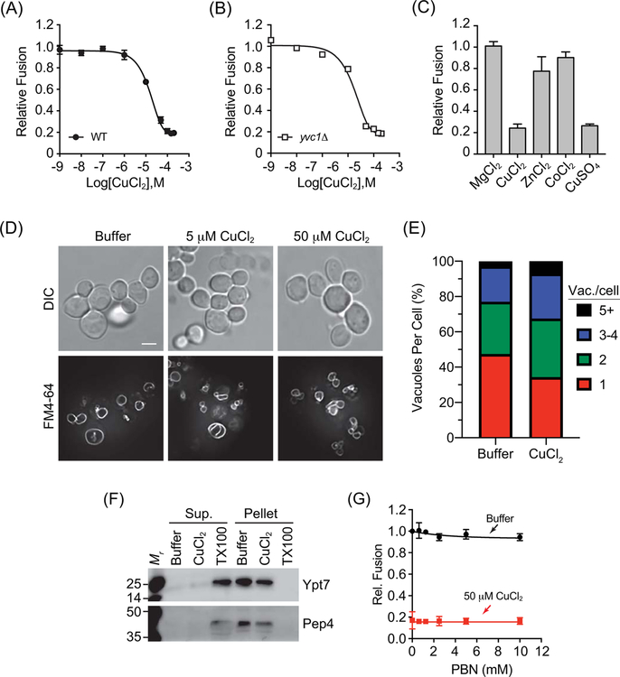
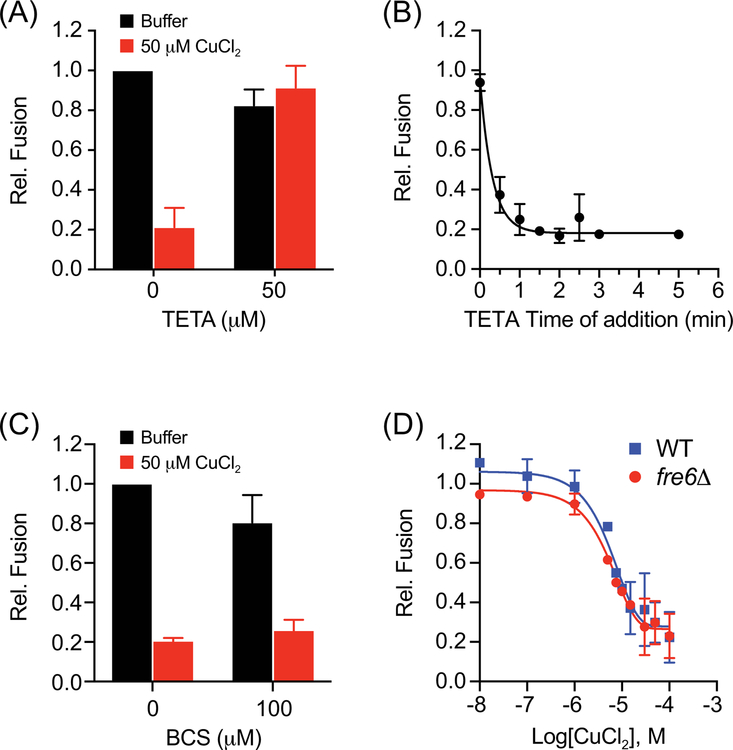
The accumulation of copper in organisms can lead to altered functions of various pathways and become cytotoxic through the generation of reactive oxygen species. In yeast, cytotoxic metals such as Hg+ , Cd2+ and Cu2+ are transported into the lumen of the vacuole through various pumps. Copper ions are initially transported into the cell by the copper transporter Ctr1 at the plasma membrane and sequestered by chaperones and other factors to prevent cellular damage by free cations. Excess copper ions can subsequently be transported into the vacuole lumen by an unknown mechanism. Transport across membranes requires the reduction of Cu2+ to Cu+ . Labile copper ions can interact with membranes to alter fluidity, lateral phase separation and fusion. Here we found that CuCl2 potently inhibited vacuole fusion by blocking SNARE pairing. This was accompanied by the inhibition of V-ATPase H+ pumping. Deletion of the vacuolar reductase Fre6 had no effect on the inhibition of fusion by copper. This suggests that Cu2+ is responsible for the inhibition of vacuole fusion and V-ATPase function. This notion is supported by the differential effects of chelators. The Cu2+ -specific chelator triethylenetetramine rescued fusion, whereas the Cu+ -specific chelator bathocuproine disulfonate had no effect on the inhibited fusion.

Keywords: Fre6; Nyv1; SNARE; Sec17; V-ATPase; Vam3; Vam7; membrane fusion.


Conflict of interest statement

CONFLICT OF INTEREST

The authors declare no potential conflict of interest.

Figures

Figure 1.
Figure 1.. CuCl2 inhibits vacuole homotypic fusion.
Vacuoles isolated from wild type (BJ3505 and DKY6281) (A) or yvc1Δ (RFY74–75) (B) fusion reporter strains were incubated with buffer alone or a concentration curve of CuCl2. (C) Wild type vacuoles were incubated with 100 μM MgCl2, ZnCl2, CoCl2, CuCl2, or CuSO4. Fusion reactions were incubated for 90 min at 27°C. After incubation membranes were solubilized and incubated with p-nitrophenyl phosphate to measure Pho8 activity. p-nitrophenolate was measured at OD400. Fusion values were normalized to the untreated control set to 1. (D) BJ3505 cells were grown to log phase and treated with CuCl2. Vacuoles were visualized by staining with FM4–64 and cells were visualized using DIC. (E) Quantitation of vacuole fragmentation of cells treated with 0 μM or 50 μM CuCl2 as in panel D. n=392, untreated cells, n=616, 50 μM CuCl2 treated cells. (F) DKY6281 vacuoles were incubated with 100 μM CuCl2, 0.1% TX-100 or buffer for 90 min at 27°C. After incubation, the soluble and membrane fractions were separated by centrifugation (16,000 x g, 10 min, 4°C), mixed with SDS-loading buffer and resolved by SDS-PAGE. The soluble luminal protease Pep4 and the membrane anchored Ypt7 were probed for by immunoblotting. (G) Wild type vacuoles were treated with or without CuCl2 in the presence of a concentration curve of PBN to eliminate oxygen radicals. Error bars are S.E.M. (n=3). Scale bar = 4 μm.
Figure 2.
Figure 2.. Cu2+ and not Cu+ inhibits vacuole fusion.
(A) Fusion reactions were incubated with or without CuCl2 and treated with TETA to chelate cupric (Cu2+) ions. (B) Fusion reactions containing 50 μM CuCl2 were supplemented with TETA at the indicated times. Fusion reactions were incubated for a total of 90 min at 27°C before developing. (C) Fusion reactions were incubated with or without CuCl2 and treated with BCS to chelate cuprous (Cu+) ions. (D) Wild type and fre6Δ vacuoles were incubated with a concentration curve of CuCl2 and tested for fusion after 90 min of incubation at 27°C. Error bars are S.E.M. (n=3).
Figure 3.
Figure 3.. Cu2+ inhibits vacuole fusion after the docking stage.
(A) Vacuoles from BJ3505 were monitored for the release of Sec17 from the membrane upon SNARE priming. Fusion reactions containing 3 μg of vacuoles (by protein) were incubated with reaction buffer, 100 μM Cu2+ or 1 mM NEM. Vacuoles were incubated at 27°C for the indicated times after witch the organelles were pelleted by centrifugation and solubilized proteins in the supernatant were separated from the membrane bound fraction. The membrane pellets were resuspended in volumes of reaction buffer equal to the supernatant. Both fractions were mixed with SDS loading buffer and resolved by SDS-PAGE. Sec17 was detected by immunoblotting and the amount released was calculated by densitometry. (B) Normalized values were averaged and plotted over time of incubation. (C) Gain of resistance kinetic vacuole fusion assays were performed in the presence of reaction buffer or 100 μM Cu2+. Reactions were incubated at 27°C or on ice for 90 min. Reagents were added at the indicated time points. Fusion inhibition was normalized to the reactions receiving buffer alone. Data were fit with first order exponential decay. Error bars are S.E.M. (n=3). (D) Isolated vacuoles harboring GFP-Ypt7 were incubated with or without 100 μM Cu2+ for 30 min at 27°C after which reaction tubes were placed on ice and labeled with FM4–64. Vacuoles were visualized by fluorescence microscopy. Scale bar = 2 μm.
Figure 4.
Figure 4.. Cu2+ inhibits SNARE complex formation.
(A) Large scale vacuole fusion reactions (6x) were incubated with anti-Sec17 IgG to block SNARE priming. After incubating for 15 min at 27°C, select reactions were further treated with either reaction buffer, 2 μM GDI or 15 μM Cu2+ and incubated for 5 min before adding 150 nM GST-Vam7. Reactions were then incubated for an additional 70 min. One reaction remained on ice for the duration of the assay. Reactions were then processed for glutathione pulldown of GST-Vam7 protein complexes. Isolated protein complexes were resolved by SDS-PAGE and probed for the SNAREs Vam3 and Nyv1. (B) Quantitation of SNARE complex formation in the presence or absence of CuCl2. Error bars are S.E.M. (n=3).
Figure 5.
Figure 5.. V-ATPase activity is blocked by Cu2+.
(A) Acridine orange (AO) fluorescence quenching assays were used to monitor H+ pumping into the vacuole lumen. Vacuoles were incubated with dose curve of CuCl2 or buffer (± ATP) and 15 μM AO. AO fluorescence quenching was measured using a plate reader. Fluorescence was measured every 40 seconds and plotted against time. (B) Quantitation of average maximum AO fluorescence. (C) AO fluorescence quenching in the presence of PS buffer (Buf.), 100 μM MgCl2, ZnCl2 or CoCl2. Error bars are S.E.M. (n=3). (D) Log phase BJ3505 cells were incubated with 50 μM CuCl2 for 1h. Vacuoles were stained with 2 μM FM4–64 and 200 μM quinacrine. DIC (Differential Interference Contrast). Scale bar = 5 μm.

References

    1. Nevitt T, Ohrvik H, Thiele DJ. Charting the travels of copper in eukaryotes from yeast to mammals. Biochim Biophys Acta. 2012;1823(9):1580–1593. - PMC - PubMed
    1. Bandmann O, Weiss KH, Kaler SG. Wilson’s disease and other neurological copper disorders. Lancet Neurol. 2015;14(1):103–113. - PMC - PubMed
    1. Cheignon C, Tomas M, Bonnefont-Rousselot D, Faller P, Hureau C, Collin F. Oxidative stress and the amyloid beta peptide in Alzheimer’s disease. Redox Biol. 2018;14450–464. - PMC - PubMed
    1. Mattson MP. Metal-catalyzed disruption of membrane protein and lipid signaling in the pathogenesis of neurodegenerative disorders. Ann N Y Acad Sci. 2004;101237–50. - PubMed
    1. Christian DA, Tian A, Ellenbroek WG, Levental I, Rajagopal K, Janmey PA, Liu AJ, Baumgart T, Discher DE. Spotted vesicles, striped micelles and Janus assemblies induced by ligand binding. Nat Mater. 2009;8(10):843–849. - PMC - PubMed

Publication types

MeSH terms

LinkOut - more resources