. 2019 Oct 1;137(10):1134-1145.
doi: 10.1001/jamaophthalmol.2019.2885.

Progression of Stargardt Disease as Determined by Fundus Autofluorescence Over a 12-Month Period: ProgStar Report No. 11

Affiliations

Progression of Stargardt Disease as Determined by Fundus Autofluorescence Over a 12-Month Period: ProgStar Report No. 11

Rupert W Strauss et al. JAMA Ophthalmol. .

Abstract

Importance: Sensitive outcome measures for disease progression are needed for treatment trials of Stargardt disease.

Objective: To estimate the progression rate of atrophic lesions in the prospective Natural History of the Progression of Atrophy Secondary to Stargardt Disease (ProgStar) study over a 12-month period.

Design, setting, and participants: This multicenter prospective cohort study was conducted in an international selection of tertiary referral centers from October 21, 2013, to February 15, 2017. Patients who were affected by Stargardt disease, aged 6 years and older at baseline, and harboring disease-causing variants of the ABCA4 gene were enrolled at 9 centers in the United States, United Kingdom, and continental Europe. Data analysis occurred from November 2016 to January 2017.

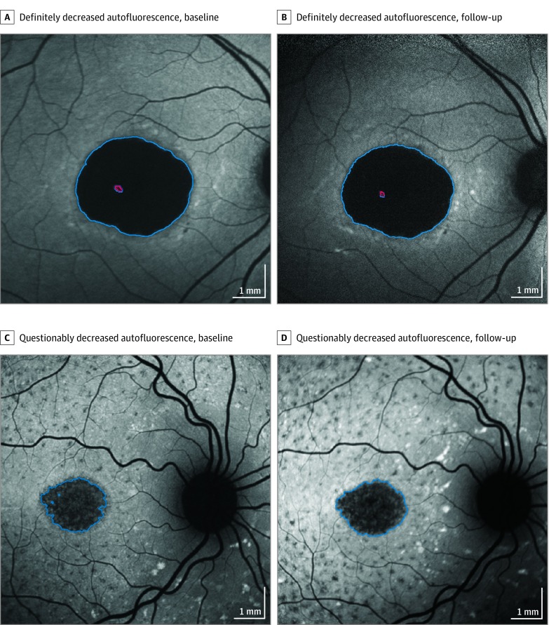
Exposures: Autofluorescence images obtained with a standard protocol were sent to a central reading center, and areas of definitely decreased autofluorescence, questionably decreased autofluorescence, and the total combined area of decreased autofluorescence were outlined and quantified. Progression rates were estimated from linear mixed models with time as the independent variable.

Main outcomes and measures: Yearly rate of progression, using the growth of atrophic lesions measured by autofluorescence imaging.

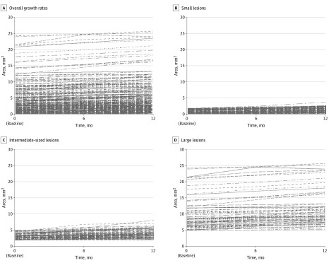
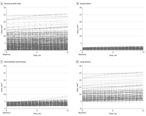
Results: A total of 259 study participants (488 eyes; 230 individuals [88.8%] were examined in both eyes) were enrolled (mean [SD] age at first visit, 33.3 [15.1] years; 118 [54.4%] female). Gradable images were available for evaluation for 480 eyes at baseline and 454 eyes after 12 months. At baseline, definitely decreased autofluorescence was present in 306 eyes, and the mean (SD) lesion size was 3.93 (4.37) mm2. The mean total area of decreased autofluorescence at baseline was 4.07 (4.04) mm2. The estimated progression of definitely decreased autofluorescence was 0.76 (95% CI, 0.54-0.97) mm2 per year (P < .001), and the total area of both questionably and definitely decreased autofluorescence was 0.64 (95% CI, 0.50-0.78) mm2 per year (P < .001). Both progression rates depended on initial lesion size.

Conclusions and relevance: In Stargardt disease, autofluorescence imaging may serve as a monitoring tool and definitely decreased autofluorescence and total area as outcome measures for interventional clinical trials that aim to slow disease progression. Rates of progression depended mainly on initial lesion size.


Conflict of interest statement

Conflict of Interest Disclosures: Dr Bernstein reports grants from the Foundation Fighting Blindness during the conduct of the study and personal fees from Spark, Acucela, Makindus, and Science Based Health, outside the submitted work. Dr Birch reports serving as a consultant for and receiving grants from Applied Genetic Technologies Corporation, Ionis, Genentech, Nightstar, 4D Molecular Therapeutics, and the Foundation Fighting Blindness; receiving personal fees from Nacuity and Editas Medicine; and receiving grants from Allergan, Second Sight Medical Products, the National Institutes of Health (grant ET09076), and the Foundation Fighting Blindness. Dr Sadda reports personal fees from Heidelberg Engineering, Centervue, Novartis, Genentech/Roche, Allergan, and Amgen; grants and personal fees from Optos; grants from Carl Zeiss Meditec; and nonfinancial support from Nidek and Topcon Medical Systems during the conduct of the study. Dr West reports membership in scientific technical advisory committee for Alcon Research Institute and Research to Prevent Blindness Inc. Dr Scholl is a paid consultant of Boehringer Ingelheim Pharma GmbH & Co, Gerson Lehrman Group, and Guidepoint; a member of the scientific advisory board of the Astellas Institute for Regenerative Medicine, ReNeuron Group plc/Ora Inc, and Vision Medicines Inc; and a member of the data monitoring and safety board/committee of Genentech Inc, Hoffmann–La Roche Ltd, and ReNeuron Group plc/Ora Inc. Dr Hendrik Scholl is a principal investigator of grants at the Universitätsspital Basel, which is sponsored by Acucela Inc, NightstaRx Ltd, and Ophthotech Corporation. Dr Strauss reports receiving stipends from the Austrian Science Fund (project number J 3383-B23) and the Foundation Fighting Blindness Clinical Research Institute. Dr Cideciyan reports receiving grants from the National Institutes of Health (grant EY013203). Dr Michaelides reports receiving career development award via the Foundation Fighting Blindness Career Development Award, the Macular Society, Retinitis Pigmentosa Fighting Blindness, Fight for Sight, and the National Institute for Health Research Biomedical Research Centre at Moorfields Eye Hospital National Health Services Foundation Trust and UCL Institute of Ophthalmology. Michael Ip reports serving as a consultant of Allergan, Thrombogenics, Omeros, Genentech, Quark, Alimera, and Boehringer Ingelheim. Dr Sahel reports grants from LabEx Lifesenses (grant ANR-10-LABX-65) and the Foundation Fighting Blindness during the conduct of the study; grants from ERC Synergy “Helmholtz” and Banque Publique d'Investissement outside the submitted work; and fees associated with work as a consultant for Pixium Vision, GenSight Biologics, and Genesignal, outside the submitted work; with additional personal financial interests in GenSight Biologics, Chronocam, Chronolife, Pixium Vision, Tilak Healthcare, and Sparing Vision. Dr Sunness reports personal fees from Acucela and Cell Cure, outside the submitted work. Dr Traboulsi reports serving on the data and safety monitoring committee for a study on gene therapy in Stargardt disease outside the submitted work. No other disclosures were reported.

Figures

Figure 1.
Figure 1.. Examples of Progression of Lesions of Definitely Decreased Autofluorescence and Questionably Decreased Autofluorescence
A, A lesion of definitely decreased autofluorescence (7.87 mm2) at baseline. B, The same eye after 12 months; the lesion is enlarged to 8.48 mm2. C, A lesion of questionably decreased autofluorescence at baseline (2.45 mm2). D, The same eye after 12 months, with the lesion enlarged to 2.90 mm2. Red indicates an excluded area without definitely decreased autofluorescence.
Figure 2.
Figure 2.. Estimated Growth Rates of Lesions of Definitely Decreased Autofluorescence
Estimated growth rates of lesions of definitely decreased autofluorescence are shown as overall growth rates (A) and subgroups of lesion size at baseline: small lesions (≤1.90 mm2) (B); intermediate-sized lesions (>1.90-5 mm2) (C), and large lesions (>5.0 mm2) (D).
Figure 3.
Figure 3.. Estimated Growth Rates of Lesions of Definitely Decreased Autofluorescence Plus Questionably Decreased Autofluorescence Combined
Overall growth rates (A), with subgroups according to lesion size at baseline: small lesions (≤1.90 mm2) (A); intermediate-sized lesions (>1.90-5 mm2) (B), and large lesions (>5.0 mm2) (C).

Comment in

  • doi: 10.1001/jamaophthalmol.2019.2930

References

    1. Allikmets R, Singh N, Sun H, et al. . A photoreceptor cell-specific ATP-binding transporter gene (ABCR) is mutated in recessive Stargardt macular dystrophy. Nat Genet. 1997;15(3):236-246. doi:10.1038/ng0397-236 - DOI - PubMed
    1. Tanna P, Strauss RW, Fujinami K, Michaelides M. Stargardt disease: clinical features, molecular genetics, animal models and therapeutic options. Br J Ophthalmol. 2017;101(1):25-30. doi:10.1136/bjophthalmol-2016-308823 - DOI - PMC - PubMed
    1. Scholl HP, Strauss RW, Singh MS, et al. . Emerging therapies for inherited retinal degeneration. Sci Transl Med. 2016;8(368):368rv6. doi:10.1126/scitranslmed.aaf2838 - DOI - PubMed
    1. Thompson DA, Ali RR, Banin E, et al. ; Monaciano Consortium . Advancing therapeutic strategies for inherited retinal degeneration: recommendations from the Monaciano Symposium. Invest Ophthalmol Vis Sci. 2015;56(2):918-931. doi:10.1167/iovs.14-16049 - DOI - PMC - PubMed
    1. Strauss RW, Ho A, Muñoz B, et al. ; Progression of Stargardt Disease Study Group . The natural history of the progression of atrophy secondary to Stargardt disease (ProgStar) studies: design and baseline characteristics, ProgStar report No. 1. Ophthalmology. 2016;123(4):817-828. doi:10.1016/j.ophtha.2015.12.009 - DOI - PubMed