. 2019 Oct;110(10):3122-3131.
doi: 10.1111/cas.14157. Epub 2019 Aug 14.

Delta-like 3 localizes to neuroendocrine cells and plays a pivotal role in gastrointestinal neuroendocrine malignancy

Affiliations

Delta-like 3 localizes to neuroendocrine cells and plays a pivotal role in gastrointestinal neuroendocrine malignancy

Kentaro Matsuo et al. Cancer Sci. 2019 Oct.

Abstract

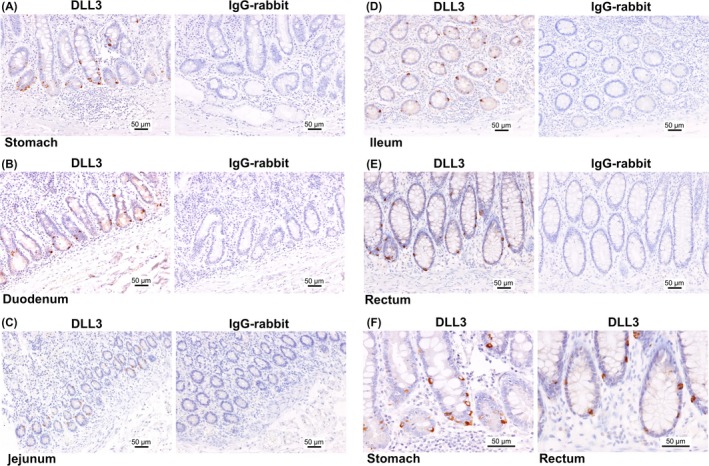
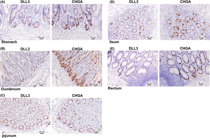
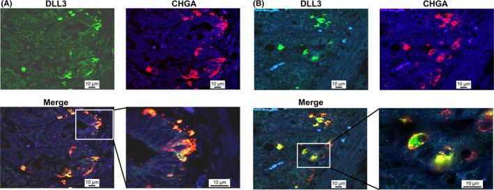
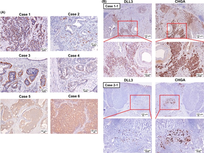
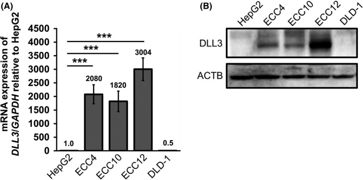
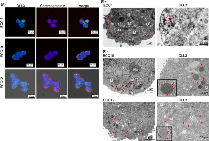
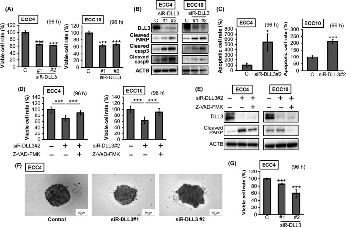
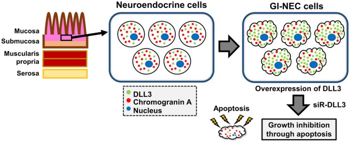
Delta-like 3 (DLL3) is a member of the Delta/Serrate/Lag2 (DSL) group of Notch receptor ligands. Five DSL ligands are known in mammals, among which DLL3 has a unique structure. In the last few years, DLL3 has attracted attention as a novel molecular targeting gene in neuroendocrine carcinoma of the lung due to its high expression. However, the expression pattern and functions of DLL3 in the gastrointestinal tract and gastrointestinal neuroendocrine carcinoma remain unclear. In this study, we examined the expression and role of DLL3 in the gastrointestinal tract, as well as in gastrointestinal neuroendocrine carcinoma. Immunohistochemical staining of the human normal gastrointestinal tract revealed that DLL3 localized in neuroendocrine cells. DLL3 showed intense staining in chromogranin A-positive gastric cancer specimens. Real-time quantitative RT-PCR and western blotting analyses showed considerable upregulation of DLL3 in gastrointestinal neuroendocrine carcinoma cell lines. Immuno-electron microscopy demonstrated abundant expression of DLL3 in neurosecretory granules in these cells. Furthermore, gene silencing of DLL3 caused significant growth inhibition through the induction of intrinsic apoptosis. Our findings suggest that DLL3 is expressed in neuroendocrine cells of the gastrointestinal tract and that it has a pivotal role in gastrointestinal neuroendocrine carcinoma cells. Based on these findings, further investigations are required to achieve a breakthrough in developing therapeutic strategies for gastrointestinal neuroendocrine carcinoma.

Keywords: DLL3; apoptosis; chromogranin A; neuroendocrine; neuroendocrine carcinoma.


Figures

Figure 1
Figure 1
The pattern of delta‐like 3 (DLL3) expression in the gastrointestinal (GI) tract. DLL3 expression in the stomach (A), duodenum (B), jejunum (C), ileum (D) and rectum (E). Left panel, the expression of DLL3; right panel, expression of IgG‐rabbit used as a negative control. F, The enlarged view of (A) and (E). Scale bar: 50 μm
Figure 2
Figure 2
The pattern of delta‐like 3 (DLL3) and chromogranin A (CHGA) expression in serial tissue sections of the GI tract. DLL3 and CHGA expression in the stomach (A), duodenum (B), jejunum (C), ileum (D) and rectum (E). Left panel, the expression of DLL3; right panel, expression of CHGA. Scale bar: 50 μm
Figure 3
Figure 3
Double immunofluorescence staining of delta‐like 3 (DLL3) and CHGA in normal stomach and duodenum. DLL3 and CHGA in the stomach (A) and duodenum (B). DLL3 is dyed green and CHGA red. Nuclei are dyed blue with DAPI. Lower panel, merged images, showing yellow fluorescence for co‐occurring antigens. White box: enlarged area of merged view. Scale bar: 10 μm
Figure 4
Figure 4
Delta‐like 3 (DLL3) expression in chromogranin A (CHGA)‐positive gastric cancer specimens. A, The expression of DLL3 in 4 CHGA‐positive gastric cancer tissue specimens and 2 GIMANEC tissue specimens. Detailed clinical information for each patient is shown in Table 1. Scale bar: 50 μm. B, The expression of DLL3 in a CHGA‐positive area of the serial tissue sections of gastric cancer specimens. The representative images of areas of cases 1 and 2 (case 1‐1 and case 2‐1) are shown. Left panel, the expression of DLL3; right panel, expression of CHGA; red box, the enlarged area. The lower panel shows the enlarged versions of each image. Scale bar: 100 μm
Figure 5
Figure 5
The expression levels of delta‐like 3 (DLL3) in gastrointestinal‐neuroendocrine carcinoma ECC cell lines. A, DLL3 mRNA expression in ECC cell lines, compared with that in other cancer cell lines (hepatocellular carcinoma cells, HepG2; and colon cancer cells, DLD‐1). The ratio of DLL3 expression to that of HepG2 was calculated, and the values are shown with HepG2 normalized to 1.0. Results are presented as the mean ± SD; ***< 0.001. B, The protein expression level of DLL3 in these cells
Figure 6
Figure 6
The morphological features of delta‐like 3 (DLL3) expression in GINEC ECC cell lines. A, The immunofluorescence staining of DLL3 and chromogranin A (CHGA) in ECC4, ECC10 and ECC12 cells. DLL3 is dyed green (left panel); CHGA, red (middle panel); merged images, showing yellow fluorescence for co‐occurring antigens (right panel). Nuclei are dyed blue with DAPI. Scale bar: 10 μm. B,C, The electron microscopy of ECC4, ECC10 and ECC12 cells. Left panel, transmission electron microscopy (TEM); right panel, immuno‐electron microscopy of anti–DLL3 antibody. Immuno‐electron microscopy for ECC4 was performed by the enzyme‐labeled antibody method and that for ECC10 and ECC12 was performed by the post–embedding immuno‐gold labeling method. G, Golgi apparatus; M, mitochondria; N, nucleus; No, nucleolus; red arrowhead, granules; red arrows, DLL3‐positive granules. Scale bar: 1.0 μm (TEM); 500 nm (immuno‐electron microscopy)
Figure 7
Figure 7
The effects of gene silencing of delta‐like 3 (DLL3) in ECC cell lines. A, Cell viability at 96 h after transfection with siR‐DLL3 (10 nmol/L). B, The protein expression levels of apoptosis‐related genes. The experimental conditions were the same as in (A). C, Results of Hoechst 33342 staining. The number of apoptotic cells was counted and shown as a bar graph. The experimental conditions were the same as in (A). D, E, Apoptosis inhibition effects of Z‐VADFMK (5 μmol/L) in ECC4 and ECC10 cells. Cells were pretreated with Z‐VADFMK and then transfected with siR‐DLL3 #2 (10 nmol/L) for 96 h. The effects on cell viability (D) and the level of cleaved PARP (E). F, G, Spheroid formation at 96 h after transfection of ECC 4 cells with siR‐DLL3 (20 nmol/L) (n = 3). Representative photographs of each sample are shown (F), and the 3D cell viabilities are shown as a bar graph (G). Results are presented as the mean ± SD; * < 0.05; *** < 0.001
Figure 8
Figure 8
Schematic diagram of the study. Delta‐like 3 (DLL3) was expressed in neuroendocrine cells of deep layer mucosa in the normal gastrointestinal (GI) tract, similar to the expression of chromogranin A (CHGA). Moreover, DLL3 expression was remarkably upregulated in neuroendocrine carcinoma (NIC) cells. Importantly, gene silencing of DLL3 caused significant growth inhibition through the induction of intrinsic apoptosis

References

    1. Radtke F, Raj K. The role of Notch in tumorigenesis: oncogene or tumour suppressor? Nat Rev Cancer. 2003;3:756‐767. - PubMed
    1. Maemura K, Yoshikawa H, Yokoyama K, et al. Delta‐like 3 is silenced by methylation and induces apoptosis in human hepatocellular carcinoma. Int J Oncol. 2013;42:817‐822. - PMC - PubMed
    1. D'Souza B, Miyamoto A, Weinmaster G. The many facets of Notch ligands. Oncogene. 2008;27:5148‐5167. - PMC - PubMed
    1. Geffers I, Serth K, Chapman G, et al. Divergent functions and distinct localization of the Notch ligands DLL1 and DLL3 in vivo. J Cell Biol. 2007;178:465‐476. - PMC - PubMed
    1. Ladi E, Nichols JT, Ge W, et al. The divergent DSL ligand Dll3 does not activate Notch signaling but cell autonomously attenuates signaling induced by other DSL ligands. J Cell Biol. 2005;170:983‐992. - PMC - PubMed

MeSH terms

Substances