. 2019 Nov 1:158:107729.
doi: 10.1016/j.neuropharm.2019.107729. Epub 2019 Jul 29.

Why flavored vape products may be attractive: Green apple tobacco flavor elicits reward-related behavior, upregulates nAChRs on VTA dopamine neurons, and alters midbrain dopamine and GABA neuron function

Affiliations

Why flavored vape products may be attractive: Green apple tobacco flavor elicits reward-related behavior, upregulates nAChRs on VTA dopamine neurons, and alters midbrain dopamine and GABA neuron function

Alicia J Avelar et al. Neuropharmacology. .

Abstract

While nicotine is the primary addictive component in tobacco products, additional flavors have become a concern with the growing popularity of electronic nicotine delivery systems (ENDS). For this reason, we have begun to investigate popular tobacco and ENDS flavors. Here, we examined farnesol, a chemical flavorant used in green apple and fruit flavors in ENDS e-liquids, for its ability to produce reward-related behavior. Using male and female 3-6 month old C57BL/6 J mice and farnesol doses of 0.1, 1, and 10 mg/kg we identified a sex-dependent effect in a conditioned place preference assay: farnesol-alone produces reward-related behavior in only male mice. Despite this sex-dependent effect, 1.0 mg/kg farnesol enhances locomotor activity in both male and female mice. To understand farnesol's effect on reward-related behavior, we used whole-cell patch-clamp electrophysiology and confocal microscopy to investigate changes in putative dopamine and GABA neurons. For these approaches, we utilized genetically modified mice that contain fluorescent nicotinic acetylcholine receptors (nAChRs). Our electrophysiological assays with male mice revealed that farnesol treatment increases ventral tegmental area (VTA) dopamine neuron firing frequency and this may be due to a decrease in inhibitory tone from GABA neurons. Our microscopy assays revealed that farnesol treatment produces a significant upregulation of α6* nAChRs in male mice but not female mice. This was supported by an observed increase in α6* nAChR function in additional electrophysiology assays. These data provide evidence that popular tobacco flavorants may alter smoking-related behavior and promote the need to examine additional ENDS flavors.

Keywords: Addiction; Conditioned place preference; Dopamine neuron; Electrophysiology; Flavorants; GABA neuron; Microscopy; Nicotinic receptors; Tobacco; Vaping.


Figures

Figure 1.
Figure 1.. Farnesol alone produces reward-related behavior in a sex-dependent manner.
(A1 and B) Male and female mice were administered saline, 0.5 mg/kg nicotine, or farnesol at doses 0.1, 1.0, or 10 mg/kg in a CPP assay. (A2) The β2* nAChR antagonist, DhβE, blocked farnesol-induced CPP in male mice. Mice failed to show significant preference for 1.0 mg/kg farnesol when given a pre-injection of 2 mg/kg DhβE. All data are mean ± SEM. *, p < 0.05; **, p < 0.01; One-way ANOVA with post hoc Bonferroni. Exact p values are given in the Results section (3.1). Number of mice for each treatment group is indicated in parenthesis.
Figure 2.
Figure 2.. 1.0 mg/kg farnesol increases nicotine conditioned place preference in only male mice.
(A and B) Male and female mice were administered saline, nicotine (0.5 mg/kg), or nicotine (0.5 mg/kg) plus farnesol (1.0 mg/kg) in a CPP assay. All data are mean ± SEM. *, p < 0.05; **, p < 0.01; One-way ANOVA with post hoc Bonferroni. Exact p values are given in the Results section (3.2). Number of mice for each treatment group is indicated in parenthesis. Individual dots within bars represent CPP scores from individual mice within the treatment groups.
Figure 3.
Figure 3.. Acute farnesol treatment increases locomotor activity in an open field.
(A - B) Immediately following an intraperitoneal injection of saline or 1.0 mg/kg farnesol, male and female mice were placed in an open field and distance traveled was measured over a duration of 20 minutes. All data are mean ± SEM. ***, p < 0.005; unpaired t-test. Exact p values are given in the Results section (3.3). The number of mice from saline- and farnesol-treated cohorts is indicated in parenthesis.
Figure 4.
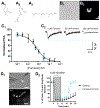
Figure 4.. Farnesol inhibits α4β2 nAChR function.
Chemical structure of menthol (A1-3) and farnesol: arranged to exhibit its similarity to menthol (A2) and its chain orientation (A3). (B1-2) Neuro-2a cells transiently transfected with α4-GFPβ2 nAChRs in brightfield (B1) and GFP (B2) imaging modes. (C1-2) α4-GFPβ2 nAChRs were stimulated with 10 μM nicotine while exposed to increasing concentrations of farnesol to examine its concentration-response on nicotine-stimulated inward currents. (C1) Mean peak current amplitude (PCA) normalized to 10 μM nicotine. Each data point represents the mean ± SEM of 5 – 7 neuro-2a cells. (C2) Representative nicotine-stimulated (10 μM, 300 ms puff) inward currents recorded from neuro-2a cells transiently transfected with α4-GFPβ2 nAChRs exposed to 0, 50, and 300 μM farnesol. Blue circles in C1 correspond to the representative waveforms in C2. (D1) Representative brightfield and GFP images of neuro-2a cells transfected with α4-GFP and β2 nAChR subunits (scale bar, 25 μm). (D2) Normalized peak current amplitudes of α4-GFPβ2 nAChRs transiently transfected in neuro-2a cells. Cells were treated with no drug (control, black) or farnesol (blue, 0.5 μM for 24 hrs). nAChRs were desensitized with a 10 minute 1 μM nicotine bath perfusion and then allowed to recover for 20 min. Before, during, and after this process, 10 μM nicotine was applied (300 ms puff at 3-minute intervals) to stimulate nAChR inward currents (n = 4 - 5 cells for each data point).
Figure 5.
Figure 5.. Farnesol treatment alters function of VTA pDA neurons.
For brain slice electrophysiology assays, male and female α6-GFP mice were treated in a manner identical to CPP assays. (A1-3) VTA pDA neurons (target bregma −3.1) were selected by the presence of α6-GFP nAChRs. (A1) Sample schematic of target VTA at bregma −3.1. (A2) Sample montage of an α6-GFP mouse brain slice at bregma −3.1. The arrow designates our target VTA pDA neurons were in the lateral as opposed to the medial regions of the VTA. Scale bars: 100 μm (A2) and 20 μm (A3). (B1-2) Representative current clamp traces of baseline firing in VTA pDA neurons from saline-treated or farnesol-treated male (B1) and female (B2) mice. (B3) Mean baseline firing frequency (mean ± SEM) for saline-treated (n = 7 neurons total from male 4 mice; 10 neurons from 4 female mice) and farnesol-treated (n = 10 neurons from 4 male mice; 10 neurons from 4 female mice). (C1) The current-voltage relationship from a series of hyperpolarizing steps to initiate Ih currents in pDA neurons of male mice (n = 4 neurons, 3 mice and 5 neurons, 4 mice for saline and farnesol-treated mice, respectively). (C2) Representative waveforms from hyperpolarizing voltage step recordings. *, p < 0.05; **, p < 0.01; unpaired t-test. Exact p values are given in the Results section (3.5).
Figure 6.
Figure 6.. Farnesol treatment decreases baseline firing frequency of putative SNr GABA neurons in male mice.
(A1) Schematic of target SNr GABA neurons at bregma −3.1. (A2) Sample images of a SNr pGABA neuron visualized in IR-DIC and fluorescence modes. All pGABA neurons recorded were negative for α6-GFP fluorescence, evidence that they are not dopamine neurons. Scale bars, 20 μm. (B1-2) Representative current clamp recordings from pGABA neurons recorded in brain slices of saline- and 1.0 mg/kg farnesol-treated male mice (treatments identical to CPP dosing paradigm). (B3) Mean firing frequency of pGABA neurons from saline-treated (n = 10 neurons, 3 mice) and farnesol-treated (n = 6 neurons, 3 mice) male mice. *, p < 0.05; unpaired t test. Exact p values are given in the Results section (3.5).
Figure 7.
Figure 7.. Acute nicotine exposure increases baseline firing frequency of putative DA and GABA neurons in male mice.
(A1-2) Representative current clamp recordings of VTA pDA neurons recorded prior to (left) and during 10 s, 500 nM nicotine application (right). (B1-2) Mean firing frequency before and during nicotine application (mean ± SEM) for saline-treated (n = 6 neurons total from 5 mice) and farnesol-treated pDA neurons (n = 5 neurons from 4 mice). (C1-2) Representative current clamp recordings of SNr pGABA neurons recorded prior to (left) and during 10 s, 500 nM nicotine application (right). (D1-2) Mean firing frequency before and during nicotine application (mean ± SEM) for saline-treated (n = 5 neurons total from 3 mice) and farnesol-treated (n = 4 neurons from 3 mice) pGABA neurons. (B3 and D3) Mean firing frequency before and during 10 s 3 μM farnesol application (mean ± SEM) for saline-treated pDA neurons (n = 4 neurons total from 3 mice) and saline-treated pGABA neurons (n = 4 neurons from 4 mice). In B1-3 and D1-3, lines indicate the individual neurons pre- and during nicotine puff. *, p < 0.05; **, p < 0.01; unpaired t-test. Exact p values are given in the Results section (3.5).
Figure 8.
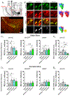
Figure 8.. Farnesol upregulates α6* and α4α6* nAChRs in VTA pDA neurons.
(A1) Schematic of our target mouse brain region in a coronal brain slice at bregma −3.1 mm. (A2) Montage of 10X images (scale bar, 100 μm) from a saline-treated α-mCherryα6-GFP mouse at bregma −3.1 mm. (B) Representative images of neurons in the VTA following saline, nicotine, nicotine plus farnesol, or farnesol-alone treatment (from CPP assays). Scale bar, 10 μm. (C1-2 and D1-2) Raw integrated density (RID) of α4*, α6*, and α4α6* nAChRs on VTA pDA neurons or RID of α4* nAChRs in SNr GABA neurons in male (C1-2) or female mice (D1-2) from saline, nicotine, nicotine plus farnesol, or farnesol-alone treatment groups. Number in parenthesis indicates the number of mice used for each treatment group. Dots within bars indicate RID values from individual mice (n = 5 and 4 for male and female mice, respectively). *, p < 0.05; **, p < 0.01; ***, p < 0.005; one-way ANOVA with post hoc Bonferroni. Exact p values are given in the Results section (3.6).
Figure 9.
Figure 9.. Long-term farnesol treatment upregulates functional α6β2β3 nAChRs in neuro-2a cells.
(A1-2 and C1-2) Representative neuro-2a cells transiently transfected with α4-GFPβ2 or α6-GFPβ2β3 nAChRs imaged in brightfield or GFP modes. Scale bars, 25 μm (B1, D1) Representative waveforms for control-treated (black) and farnesol-treated (blue) α4-GFPβ2 nAChRs (B1) and α6-GFPβ2β3 nAChRs (D1). (B2 and D2) Mean peak current amplitude of 300 ms puffs of 10 μM nicotine. Arrows indicate start of 300 ms puff. Data are mean ± SEM and individual dots represent individual cells (n = 5 - 6 cells, each condition). Exact p values are given in the Results section (3.7).
Figure 10.
Figure 10.. Summary of farnesol’s effect on midbrain neurons.
VTA dopamine neurons receive excitatory inputs from local/distant glutamate (Glu) neurons and cholinergic neurons but inhibitory input from GABA neurons. Compared to saline-treated dopamine neurons (A), farnesol-treated neurons (B) exhibit an increase in α4α6* and α6* nAChRs and elevated action potential frequency. This elevation in dopamine neuron firing frequency is likely a consequence of decreased activity from inhibitory GABA neurons. PPtg, pedunculopontine tegmental nucleus; LDTg, laterodorsal tegmental nucleus.

References

    1. Alsharari SD, King JR, Nordman JC, Muldoon PP, Jackson A, Zhu AZX, Tyndale RF, Kabbani N, Damaj MI (2015) Effects of menthol on nicotine pharmacokinetic, pharmacology and dependence in mice. PLoS One 10:e0137070. - PMC - PubMed
    1. Antoniou K, Papathanasiou G, Papalexi E, Hyphantis T, Nomikos GG, Spyraki C, Papadopoulou-Daifoti Z (2008) Individual responses to novelty are associated with differences in behavioral and neurochemical profiles. Behav Brain Res 187:462–472. - PubMed
    1. Aszyk J, Kubica P, Wozniak MK, Namiesnik J, Wasik A, Kot-Wasik A (2018) Evaluation of flavour profiles in e-cigarette refill solutions using gas chromatography-tandem mass spectrometry. J Chromatogr A 1547:86–98. - PubMed
    1. Baker LK, Mao D, Chi H, Govind AP, Vallejo YF, Iacoviello M, Herrera S, Cortright JJ, Green WN, McGehee DS, Vezina P (2013) Intermittent nicotine exposure upregulates nAChRs in VTA dopamine neurons and sensitises locomotor responding to the drug. Eur J Neurosci 37:1004–1011. - PMC - PubMed
    1. Benowitz NL, Samet JM (2011) The threat of menthol cigarettes to U.S. public health. N Engl J Med 364:2179–2181. - PubMed

Publication types

MeSH terms

LinkOut - more resources