Activin A-mediated epithelial de-differentiation contributes to injury repair in an in vitro gastrointestinal reflux model
- PMID: 31369967
- PMCID: PMC6739184
- DOI: 10.1016/j.cyto.2019.154782
Activin A-mediated epithelial de-differentiation contributes to injury repair in an in vitro gastrointestinal reflux model
Abstract
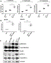
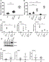
Reflux esophagitis is a result of esophageal exposure to acid and bile during episodes of gastroesophageal reflux. Aside from chemical injury to the esophageal epithelium, it has been shown that acid and bile induce cytokine-mediated injury by stimulating the release of pro-inflammatory cytokines. During the repair and healing process following reflux injury, the squamous esophageal cells are replaced with a columnar epithelium causing Barrett's metaplasia, which predisposes patients to esophageal adenocarcinoma. We identified a novel player in gastroesophageal reflux injury, the TGFβ family member Activin A (ActA), which is a known regulator of inflammation and tissue repair. In this study, we show that in response to bile salt and acidified media (pH 4) exposure, emulating the milieu to which the distal esophagus is exposed during gastroesophageal reflux, long-term treated, tolerant esophageal keratinocytes exhibit increased ActA secretion and a pro-inflammatory cytokine signature. Furthermore, we noted increased motility and expression of the stem cell markers SOX9, LGR5 and DCLK1 supporting the notion that repair mechanisms were activated in the bile salt/acid-tolerant keratinocytes. Additionally, these experiments demonstrated that de-differentiation as characterized by the induction of YAP1, FOXO3 and KRT17 was altered by ActA/TGFβ signaling. Collectively, our results suggest a pivotal role for ActA in the inflammatory GERD environment by modulating esophageal tissue repair and de-differentiation.
Keywords: Barrett’s esophagus; Gastroesophageal reflux; Inflammation; Stem cell markers; TGFβ signaling.
Copyright © 2019 Elsevier Ltd. All rights reserved.
Figures


Similar articles
-
Acidic Bile Salts Induce Epithelial to Mesenchymal Transition via VEGF Signaling in Non-Neoplastic Barrett's Cells.Gastroenterology. 2019 Jan;156(1):130-144.e10. doi: 10.1053/j.gastro.2018.09.046. Epub 2018 Sep 27. Gastroenterology. 2019. PMID: 30268789 Free PMC article.
-
Barrett's metaplasia develops from cellular reprograming of esophageal squamous epithelium due to gastroesophageal reflux.Am J Physiol Gastrointest Liver Physiol. 2017 Jun 1;312(6):G615-G622. doi: 10.1152/ajpgi.00268.2016. Epub 2017 Mar 23. Am J Physiol Gastrointest Liver Physiol. 2017. PMID: 28336546
-
From Reflux Esophagitis to Esophageal Adenocarcinoma.Dig Dis. 2016;34(5):483-90. doi: 10.1159/000445225. Epub 2016 Jun 22. Dig Dis. 2016. PMID: 27331918 Free PMC article. Review.
-
Factors influencing the development of Barrett's epithelium in the esophageal remnant postesophagectomy.Am J Gastroenterol. 2004 Feb;99(2):205-11. doi: 10.1111/j.1572-0241.2004.04057.x. Am J Gastroenterol. 2004. PMID: 15046206
-
Reflux esophagitis and its role in the pathogenesis of Barrett's metaplasia.J Gastroenterol. 2017 Jul;52(7):767-776. doi: 10.1007/s00535-017-1342-1. Epub 2017 Apr 27. J Gastroenterol. 2017. PMID: 28451845 Free PMC article. Review.
Cited by
-
Identification of Unique Transcriptomic Signatures and Hub Genes Through RNA Sequencing and Integrated WGCNA and PPI Network Analysis in Nonerosive Reflux Disease.J Inflamm Res. 2021 Nov 23;14:6143-6156. doi: 10.2147/JIR.S340452. eCollection 2021. J Inflamm Res. 2021. PMID: 34848992 Free PMC article.
-
Assessment of the risk factors of duodenogastric reflux in relation to different dietary habits in a Chinese population of the Zhangjiakou area.Food Nutr Res. 2023 Oct 24;67. doi: 10.29219/fnr.v67.9385. eCollection 2023. Food Nutr Res. 2023. PMID: 37920676 Free PMC article.
-
Serum activin A as a prognostic biomarker for community acquired pneumonia.World J Clin Cases. 2024 Aug 6;12(22):5016-5023. doi: 10.12998/wjcc.v12.i22.5016. World J Clin Cases. 2024. PMID: 39109010 Free PMC article. Clinical Trial.
-
Mechanisms and pathophysiology of Barrett oesophagus.Nat Rev Gastroenterol Hepatol. 2022 Sep;19(9):605-620. doi: 10.1038/s41575-022-00622-w. Epub 2022 Jun 7. Nat Rev Gastroenterol Hepatol. 2022. PMID: 35672395 Review.
-
Novel risk loci encompassing genes influencing STAT3, GPCR, and oxidative stress signaling are associated with co-morbid GERD and COPD.PLoS Genet. 2025 Feb 7;21(2):e1011531. doi: 10.1371/journal.pgen.1011531. eCollection 2025 Feb. PLoS Genet. 2025. PMID: 39919125 Free PMC article.
References
-
- Shaheen NJ, Hansen RA, Morgan DR, Gangarosa LM, Ringel Y, Thiny MT, Russo MW, Sandler RS, The burden of gastrointestinal and liver diseases, 2006, The American journal of gastroenterology 101(9) (2006) 2128–38. - PubMed
-
- Spechler SJ, Souza RF, Barrett’s esophagus, The New England journal of medicine 371(9) (2014) 836–45. - PubMed
-
- Pham TH, Genta RM, Spechler SJ, Souza RF, Wang DH, Development and characterization of a surgical mouse model of reflux esophagitis and Barrett’s esophagus, Journal of gastrointestinal surgery: official journal of the Society for Surgery of the Alimentary Tract 18(2) (2014) 234–40; discussion 240–1. - PubMed
Publication types
MeSH terms
Substances
Grants and funding
LinkOut - more resources
Full Text Sources
Medical
Research Materials
Miscellaneous

