. 2019 Jul 31;20(15):3731.
doi: 10.3390/ijms20153731.

Molecular and Hormonal Aspects of Drought-Triggered Flower Shedding in Yellow Lupine

Affiliations

Molecular and Hormonal Aspects of Drought-Triggered Flower Shedding in Yellow Lupine

Emilia Wilmowicz et al. Int J Mol Sci. .

Abstract

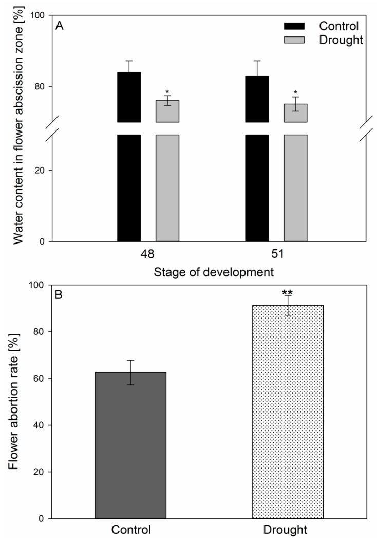
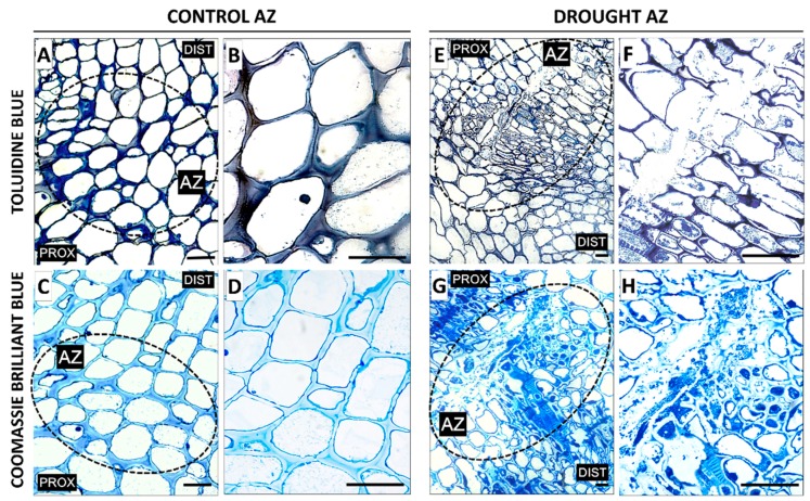
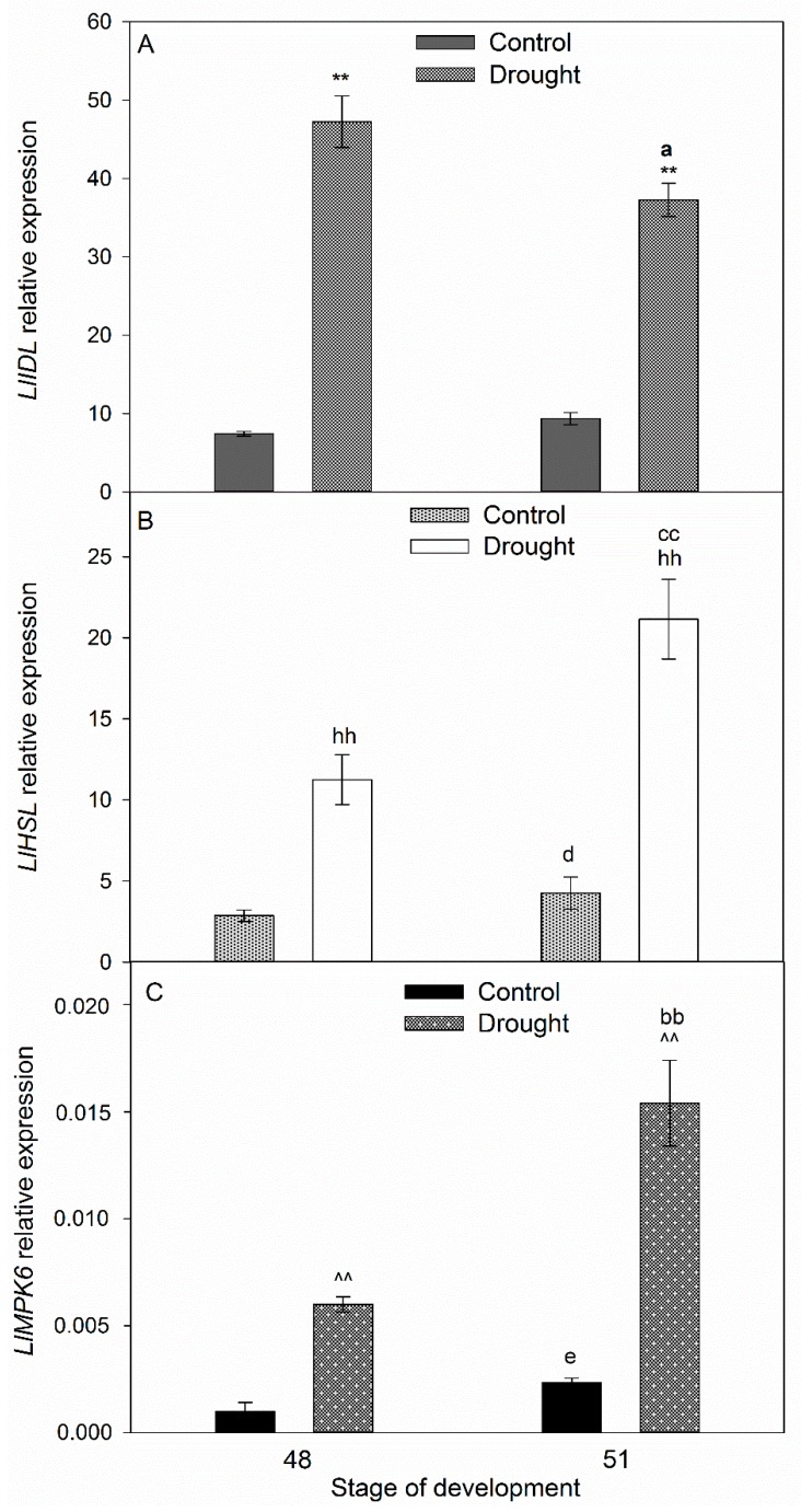
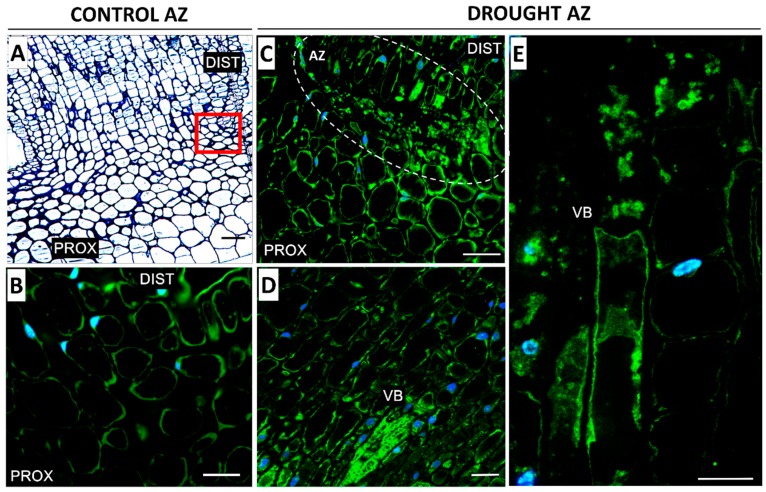
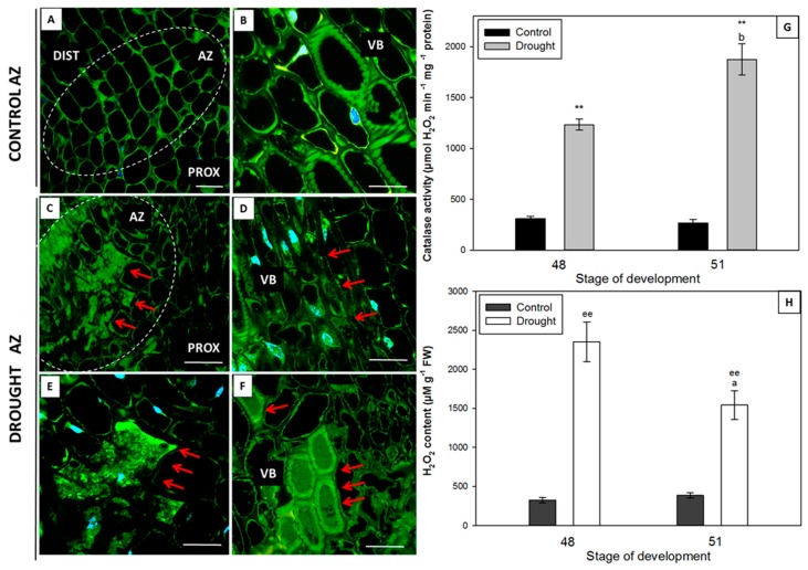
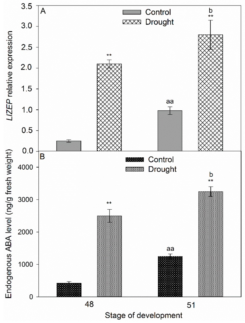
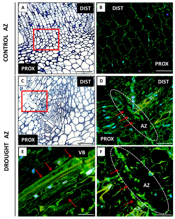
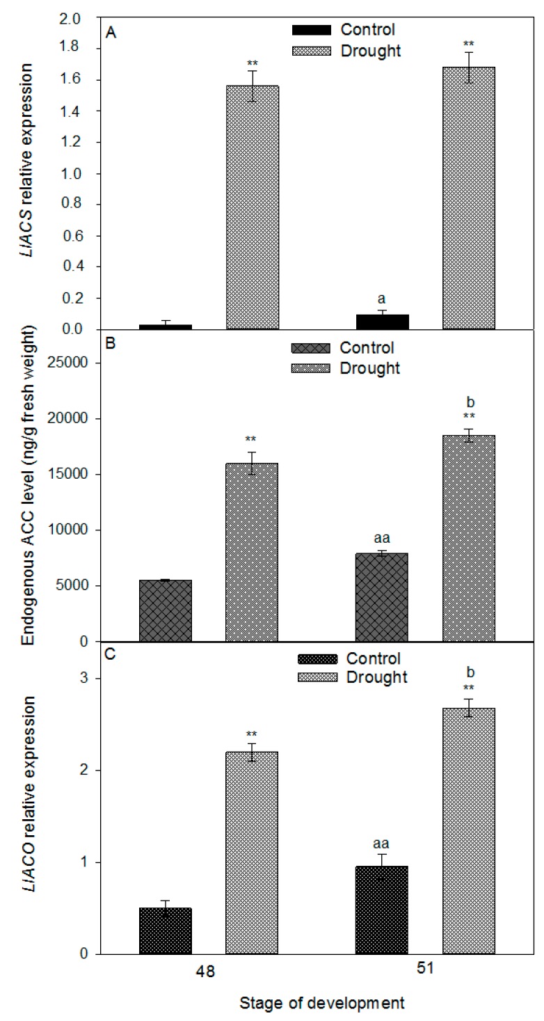
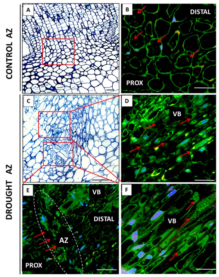
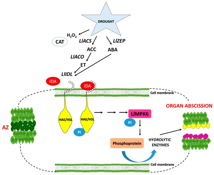
The drought is a crucial environmental factor that determines yielding of many crop species, e.g., Fabaceae, which are a source of valuable proteins for food and feed. Herein, we focused on the events accompanying drought-induced activation of flower abscission zone (AZ)-the structure responsible for flower detachment and, consequently, determining seed production in Lupinus luteus. Therefore, detection of molecular markers regulating this process is an excellent tool in the development of improved drought-resistant cultivars to minimize yield loss. We applied physiological, molecular, biochemical, immunocytochemical, and chromatography methods for a comprehensive examination of changes evoked by drought in the AZ cells. This factor led to significant cellular changes and activated AZ, which consequently increased the flower abortion rate. Simultaneously, drought caused an accumulation of mRNA of genes inflorescence deficient in abscission-like (LlIDL), receptor-like protein kinase HSL (LlHSL), and mitogen-activated protein kinase6 (LlMPK6), encoding succeeding elements of AZ activation pathway. The content of hydrogen peroxide (H2O2), catalase activity, and localization significantly changed which confirmed the appearance of stressful conditions and indicated modifications in the redox balance. Loss of water enhanced transcriptional activity of the abscisic acid (ABA) and ethylene (ET) biosynthesis pathways, which was manifested by elevated expression of zeaxanthin epoxidase (LlZEP), aminocyclopropane-1-carboxylic acid synthase (LlACS), and aminocyclopropane-1-carboxylic acid oxidase (LlACO) genes. Accordingly, both ABA and ET precursors were highly abundant in AZ cells. Our study provides information about several new potential markers of early response on water loss, which can help to elucidate the mechanisms that control plant response to drought, and gives a useful basis for breeders and agronomists to enhance tolerance of crops against the stress.

Keywords: LlHSL; LlIDA; LlMPK6; abscisic acid; abscission zone; catalase; drought stress; ethylene; hormone homeostasis; yellow lupine.


Conflict of interest statement

The authors declare no conflict of interest.

Figures

Figure 1
Figure 1
The influence of soil drought stress on the water content in flower abscission zone (AZ) [%] (A) and flower abortion rate (%) (B) in Lupinus luteus. Control plants were growing in soil of optimal moisture (70% holding capacity; WHC). In parallel, part of plants was subjected to drought conditions for 2 weeks (25% WHC). Plant material for water content measurements was collected on the 48th and 51st days of cultivation (A). The number of aborted flowers per one lupine was counted and the data were presented as averages of 15 technical replicates ± SE. Significant differences to stress plants in comparison to control are indicated as * p < 0,05, ** p < 0.01 (Student’s t-test).
Figure 2
Figure 2
The impact of soil drought stress (25% WHC) on the structural features in the flower abscission zone (AZ) of Lupinus luteus. For observations, sections of AZ were excised on the 48th day of cultivation. Samples of AZ from control and drought-stressed plants were stained with toluidine blue (A,B,E,F) or Coomassie Brilliant Blue (C,D,G,H). Images B, D, F, and H are magnifications of different areas presented in A, C, E, G, respectively. AZ area was marked by black dotted circle (A,C,E,G). Abbreviations: PROX—stem fragments below the AZ, DIST—flower pedicel fragments above the AZ. Scale bars: 40 µm.
Figure 3
Figure 3
Transcriptional activity of LlIDL (A), LlHSL (B), and LlMPK6 (C) (related to LlACT) in the yellow lupine flower abscission zone (AZ) subjected to drought. Control plants were growing in soil of optimal moisture (70% WHC), while part of plants was subjected to drought conditions for 2 weeks (25% WHC). For gene expression profiling, AZs were harvested on the 48th and 51st days of cultivation. Data are presented as averages ± SE. For LlIDL expression, significant differences in stressed plants in comparison to control plants are indicated as ** p < 0.01, and significant differences in 51-day-old stressed plants in comparison to 48-day-old plants are indicated as a p < 0.05. For LlHSL, significant differences in stressed plants in comparison to control plants are indicated as hh p < 0.01, significant differences in 51-day-old control plants in comparison to 48-day-old plants are indicated as d p < 0.05, and significant differences in 51-day-old stressed plants in comparison to 48-day-old stressed plant are indicated as cc p < 0.01. For LlMPK6, significant differences in stressed plants in comparison to control plants are indicated as ^^ p < 0.01, significant differences in 51-day-old control plants in comparison to 48-day-old plants are indicated as e p < 0.05, and significant differences in 51-day-old stressed plants in comparison to 48-day-old stressed plants are indicated as bb p < 0.01 (Student’s t-test).
Figure 4
Figure 4
Immunofluorescence localization of MPK6 in the abscission zone (AZ) of yellow lupine flowers grown under drought conditions (CE) and in the control AZ (B). The AZs were excised on the 48th day of development. Control plants were cultivated under optimal soil conditions (70% WHC), whereas stressed plants were subjected to drought for 2 weeks (25% WHC). The AZ region of stressed plants was highlighted by white lines (C). Images D and E present the vascular bundle area. Nuclei were stained with DAPI. The examined region of control AZ used in the immunofluorescence studies os indicated by a red square (A). Abbreviations: PROX—stem fragments below the AZ, DIST—flower pedicel fragments above the AZ, VB—vascular bundles. Scale bars: 100 µm (A, C), 40 µm (B,D,E).
Figure 5
Figure 5
Immunolocalization of catalase (CAT) in the floral abscission zone (AZ) of yellow lupine grown in drought conditions (CF) and in the AZ of control plants (A,B). The AZs were excised on the 48th day of development. Control plants were cultivated under optimal soil conditions (70% WHC). Stressed plants were subjected to drought for 2 weeks (25% WHC). The AZ region is indicated by white curves (A,C). The presence of CAT is highlighted by red arrows. Image E is magnified C. Nearby cells to vascular bundles (D). Magnified vascular bundles (B,F). Nuclei were stained with DAPI. Abbreviations: PROX—stem fragments below the AZ, DIST—flower pedicel fragments above the AZ, VB—vascular bundles. Scale bars: 100 µm (A,C), 60 µm (D,F), 40 µm (B,E). CAT activity (G) and hydrogen peroxide (H2O2) concentration (H) in the floral AZ of yellow lupine grown in drought conditions and in the AZ of control plants. Data are presented as averages ± SE. For CAT activity, significant differences in stressed plants in comparison to control plants are indicated as ** p < 0.01, and in 51-day-old stressed plants in comparison to 48-day-old plants are indicated as b p < 0.05. For H2O2 content, significant differences in stressed plants in comparison to control plants are indicated as ee p < 0.01, and in 51-day-old stressed plants in comparison to 48-day-old plants are indicated as a p < 0.05.
Figure 6
Figure 6
LlZEP expression (related to LlACT) (A) and endogenous content of abscisic acid (ABA) in floral abscission zone (AZ) of Lupinus luteus grown under drought conditions. Control plants were growing in soil of optimal moisture (70% WHC). In parallel, part of plants was subjected to drought for 2 weeks (25% WHC). For analysis, AZs were harvested on the 48th and 51st days of cultivation. Data are presented as averages ± SE. For LlZEP expression, significant differences in stressed plants in comparison to control plants are indicated as ** p < 0.01, significant differences in 51-day-old control plants in comparison to 48-day-old plants are indicated as aa p < 0.01, and significant differences in 51-day-old stressed plants in comparison to 48-day-old stressed plant are indicated as b p < 0.05. For ABA content, significant differences in the stressed plants in comparison to control plants are indicated as ** p < 0.01, significant differences in 51-day-old control plants in comparison to 48-day-old plants are indicated as aa p < 0.01, and significant differences in 51-day-old stressed plants in comparison to 48-day-old stressed plants are indicated as b p < 0.05 (Student’s t-test).
Figure 7
Figure 7
Tissue and subcellular localization of abscisic acid (ABA) in the abscission zone (AZ) of Lupinus luteus flowers grown in drought conditions (DF) and in the AZ of control plants (B). The AZs were excised on the 48th day of development. Control plants were cultivated under optimal soil conditions (70% WHC). Part of plants was subjected to drought conditions for 2 weeks (25% WHC). The AZ regions are indicated by white curves (D,F). The presence of ABA (DF) is highlighted by red arrows. Image F is magnified D region. Image E corresponds to the vascular bundles’ area. DAPI was used for nuclei staining. The examined regions of AZs used for the immunofluorescence studies are indicated by red squares (A,C). Abbreviations: PROX—stem fragments below the AZ, DIST—flower pedicel fragments above the AZ, VB—vascular bundles. Scale bars: 100 µm (A,C,D), 60 µm (B), 40 µm (E,F).
Figure 8
Figure 8
Expression analysis of ethylene (ET) biosynthesis genes, LlACS (A), and LlACO (C), (related to LlACT) and endogenous level of ET precursor-ACC in floral abscission zone (AZ) of Lupinus luteus grown under drought conditions. Control plants were cultivated in soil of optimal moisture (70% WHC). Part of plants was subjected to drought conditions for 2 weeks (25% WHC). AZs were harvested on the 48th and 51st days of cultivation. Data are presented as averages ± SE. For LlACS expression, significant differences in stressed plants in comparison to control plants are indicated as ** p < 0.01, and significant differences in 51-day-old control plants in comparison to 48-day-old plants are indicated as a p < 0.05. For LlACO expression, significant differences in stressed plants in comparison to control plants are indicated as ** p < 0.01, significant differences in 51-day-old control plants in comparison to 48-day-old plants are indicated as aa p < 0.01, and significant differences in 51-day-old stressed plants in comparison to 48-day-old stressed plant are indicated as b p < 0.05. For ACC content, significant differences in stressed plants in comparison to control plants are indicated as ** p < 0.01, significant differences in 51-day-old control plants in comparison to 48-day-old plants are indicated as aa p < 0.01, and significant differences in 51-day-old stressed plants in comparison to 48-day-old stressed plant are indicated as b p < 0.05 (Student’s t-test).
Figure 9
Figure 9
Immunolocalization of ethylene precursor, ACC, in the floral abscission zone (AZ) of yellow lupine grown in drought conditions (D, E, F) and in the AZ of control plants (B). The AZs were excised on the 48th day of development. Control plants were cultivated under optimal soil conditions (70% WHC), part of plants was subjected to drought conditions for 2 weeks (25% WHC). The AZ region is highlighted by white curves (E). The presence of ACC is indicated by red arrows. D and F correspond to vascular bundle areas. Nuclei were stained with DAPI. The studied regions of AZs used for the immunofluorescence studies are marked by red squares (A,C). Abbreviations: PROX—stem fragments below the AZ, DIST—flower pedicel fragments above the AZ, VB—vascular bundles. Scale bars: 100 µm (A,C), 60 µm (D,E), 40 µm (B,F).
Figure 10
Figure 10
Hypothetical model of floral abscission zone functioning under drought stress in yellow lupine. The precise description is in the text.

Similar articles

Cited by

References

    1. McKeown A.W., Warland J., McDonald M.R. Long-term climate and weather patterns in relation to crop yield: A mini review. Can. J. Bot. 2006;84:1031–1037. doi: 10.1139/b06-080. - DOI
    1. Lucas M.M., Stoddard F.L., Annicchiarico P., Frías J., Martínez-Villaluenga C., Sussmann D., Duranti M., Seger A., Zander P.M., Pueyo J.J. The future of lupin as a protein crop in Europe. Front. Plant Sci. 2015;6:705. doi: 10.3389/fpls.2015.00705. - DOI - PMC - PubMed
    1. Ainouche A.K., Bayer R.J., Misset M.T. Molecular phylogeny, diversification and character evolution in Lupinus (Fabaceae) with special attention to Mediterranean and African lupines. Plant Syst. Evol. 2004;246:211–222. doi: 10.1007/s00606-004-0149-8. - DOI
    1. Estornell L.H., Agustí J., Merelo P., Talón M., Tadeo F.R. Elucidating mechanisms underlying organ abscission. Plant Sci. 2013;199:48–60. doi: 10.1016/j.plantsci.2012.10.008. - DOI - PubMed
    1. Wilmowicz E., Frankowski K., Kućko A., Świdziński M., de Dios Alché J., Nowakowska A., Kopcewicz J. The influence of abscisic acid on the ethylene biosynthesis pathway in the functioning of the flower abscission zone in Lupinus luteus. J. Plant Physiol. 2016;206:49–58. doi: 10.1016/j.jplph.2016.08.018. - DOI - PubMed

MeSH terms

LinkOut - more resources