. 2019 Jul 31;20(15):3732.
doi: 10.3390/ijms20153732.

A Novel Adenosine Kinase from Bombyx mori: Enzymatic Activity, Structure, and Biological Function

Affiliations

A Novel Adenosine Kinase from Bombyx mori: Enzymatic Activity, Structure, and Biological Function

Kai Song et al. Int J Mol Sci. .

Abstract

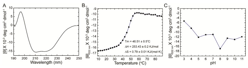
Adenosine kinase (ADK) is the first enzyme in the adenosine remediation pathway that catalyzes adenosine phosphorylation into adenosine monophosphate, thus regulating adenosine homeostasis in cells. To obtain new insights into ADK from Bombyx mori (BmADK), we obtained recombinant BmADK, and analyzed its activity, structure, and function. Gel-filtration showed BmADK was a monomer with molecular weight of approximately 38 kDa. Circular dichroism spectra indicated BmADK had 36.8% α-helix and 29.9% β-strand structures, respectively. The structure of BmADK was stable in pH 5.0-11.0, and not affected under 30 °C. The melting temperature and the enthalpy and entropy changes in the thermal transition of BmADK were 46.51 ± 0.50 °C, 253.43 ± 0.20 KJ/mol, and 0.79 ± 0.01 KJ/(mol·K), respectively. Site-directed mutagenesis demonstrated G68, S201, E229, and D303 were key amino acids for BmADK structure and activity. In particular, S201A mutation significantly increased the α-helix content of BmADK and its activity. BmADK was located in the cytoplasm and highly expressed in the silk gland during the pre-pupal stage. RNA interference revealed the downregulation of BmADK decreased ATG-8, Caspase-9, Ec-R, E74A, and Br-C expression, indicating it was likely involved in 20E signaling, apoptosis, and autophagy to regulate silk gland degeneration and silkworm metamorphosis. Our study greatly expanded the knowledge on the activity, structure, and role of ADK.

Keywords: Bombyx mori; adenosine kinase; enzymatic activity; structure.


Conflict of interest statement

All authors declare there are no conflicts of interest, including specific financial interests and relationships and affiliations relevant to the subject of this manuscript.

Figures

Figure 1
Figure 1
Structural prediction, purification of heterologously expressed Bombyx mori adenosine kinase (BmADK), and sequence comparison of ADK from different species. (A) Diagram shows the predicted secondary structure of BmADK. (B) Purification of BmADK by gel filtration and SDS-PAGE analysis. (C) Multiple sequence alignments of ADKs from Bombyx mori, Amyelois transitella, Papilio troilus, Stomoxys calcitrans, Lucilia cuprina, Nicrophorus orbicollis, and Cephus cinctus. Similarities are highlighted in red and mutation sites are marked with blue stars.
Figure 2
Figure 2
Secondary structure and stability analysis of BmADK. (A) CD spectra of BmADK. (B) Structural changes of BmADK induced by temperature. (C) Structural changes of BmADK in pH range of 3.0–11.0. The mean residue ellipticities at 222 nm are used to represent the structural changes of BmADK induced by pH and temperature.
Figure 3
Figure 3
Influence of mutations on BmADK activity and secondary structure. (A) Determination of enzymatic activity of BmADK mutants and WT. (B) Comparison of enzymatic activity between BmADK mutants and WT. All experiments were performed in triplicate. Data are represented as mean ± SD. ***, p < 0.001, compared to the control. (C,D) CD spectra of BmADK and its mutants. The mean residue ellipticity was the arithmetic mean of three independent tests. (E) Homologous modeling of the substrate-binding pocket of BmADK. The catalytic residues (S201, E229, D303) are shown in sticks.
Figure 4
Figure 4
Expression profile and cellular location of BmADK. (A) Expression profile of BmADK in the head, malpighian tubules, gonad, midgut, fat body, silk gland, blood cell, and epidermis of the silkworm on the third day of the fifth instar. (B) Expression profile of BmADK in the silk gland at mRNA level from the first day of the fifth instar to the first day of the pupa of the silkworm. 5th, the fifth instar stage; W, wandering stage; Pupa, pupal stage. (C) Fluorescence location of BmADK in BmE cells. “Red” is the signal from recombinant BmADK. The cells were stained with DAPI for 20 min. Scale bar is 20 μm.
Figure 5
Figure 5
Effects of BmADK RNAi on cell apoptosis, autophagy, and 20E signaling. (A) BmADK expression at 36 h after RNAi with ds-BmADK. The expression of (B) ATG 8, (C) Caspase-9, (D) Ec-R, (E) E74A, and (F) Br-C at 36 h after RNAi with ds-BmADK. EGFP was used as the control. All experiments were performed in triplicate. Data are represented as mean ± SD. *, p < 0.05; **, p < 0.01; ***, p < 0.001, compared to the control.

Similar articles

Cited by

References

    1. Krenitsky T.A., Miller R.L., Fyfe J.A. Levels of nucleoside and nucleotide kinases in Rhesus monkey tissues. Biochem. Pharmacol. 1974;23:170–172. doi: 10.1016/0006-2952(74)90325-6. - DOI - PubMed
    1. Snyder F.F., Lukey T. Kinetic considerations for the regulation of adenosine and deoxyadenosine metabolism in mouse and human tissues based on a thymocyte model. Biochim. Biophys. Acta. 1982;696:299–307. doi: 10.1016/0167-4781(82)90061-6. - DOI - PubMed
    1. Singh B., Hao W.H., Wu Z.C., Eigl B., Gupta R.S. Cloning and characterization of cDNA for adenosine kinase from mammalian (Chinese hamster, mouse, human and rat) species: High frequency mutants of Chinese hamster ovary cells involve structural alterations in the gene. Eur. J. Biochem. 1996;241:564–571. doi: 10.1111/j.1432-1033.1996.00564.x. - DOI - PubMed
    1. Spychala J., Datta N.S., Takabayashi K., Datta M., Fox I.H., Gribbin T., Mitchell B.S. Cloning of human adenosine kinase cDNA: Sequence similarity to microbial ribokinases and fructokinases. Proc. Natl. Acad. Sci. USA. 1996;93:1232–1237. doi: 10.1073/pnas.93.3.1232. - DOI - PMC - PubMed
    1. Decking U.K.M., Schlieper G., Kroll K., Schrader J. Hypoxia-induced inhibition of adenosine kinase potentiates cardiac adenosine release. Circ. Res. 1997;81:154–164. doi: 10.1161/01.RES.81.2.154. - DOI - PubMed

MeSH terms

LinkOut - more resources