Review
. 2019 Jul 31;8(8):805.
doi: 10.3390/cells8080805.

Metabolic Plasticity of Acute Myeloid Leukemia

Affiliations
Review

Metabolic Plasticity of Acute Myeloid Leukemia

Johanna Kreitz et al. Cells. .

Abstract

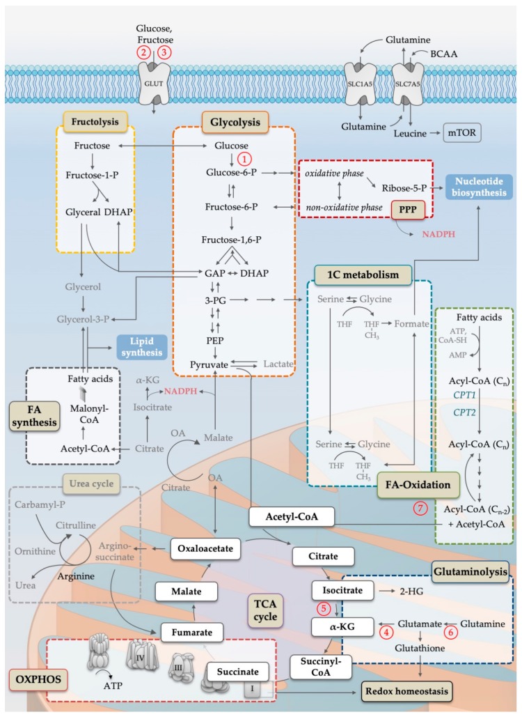
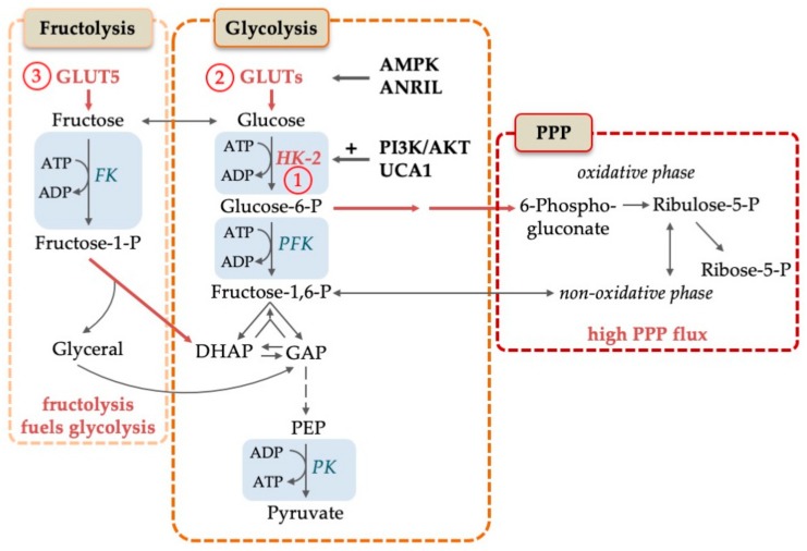
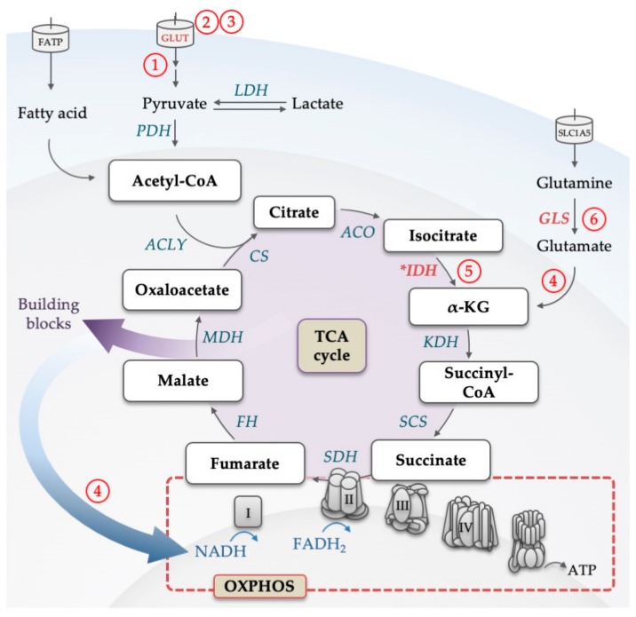
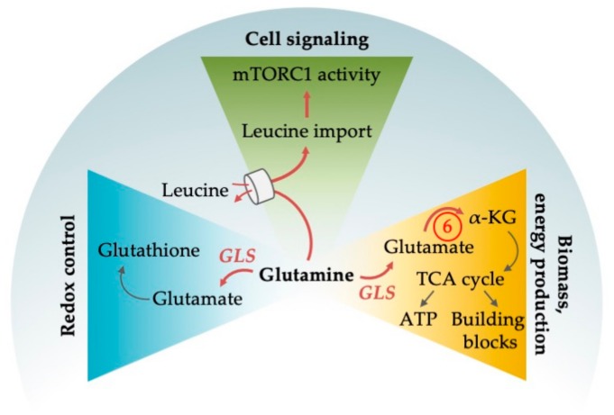
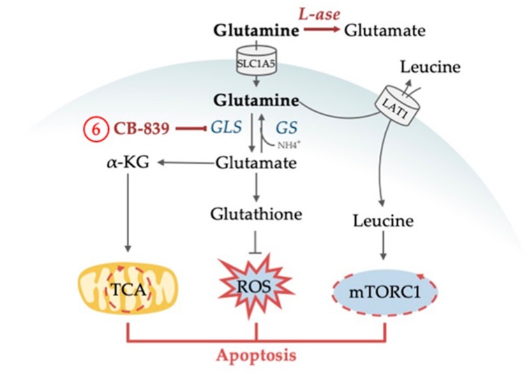
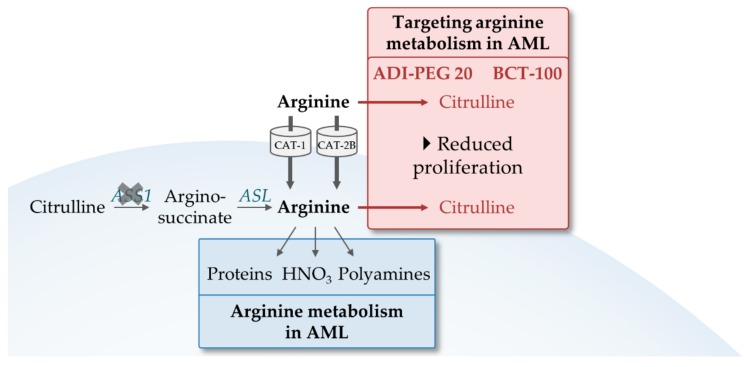
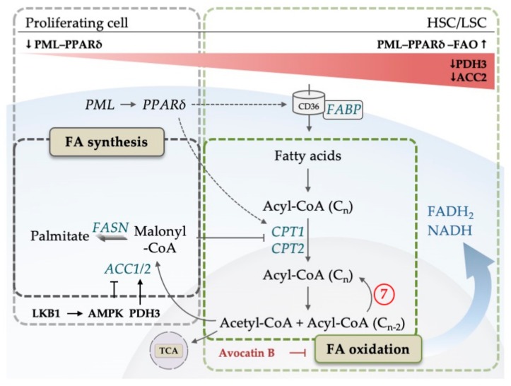
Acute myeloid leukemia (AML) is one of the most common and life-threatening leukemias. A highly diverse and flexible metabolism contributes to the aggressiveness of the disease that is still difficult to treat. By using different sources of nutrients for energy and biomass supply, AML cells gain metabolic plasticity and rapidly outcompete normal hematopoietic cells. This review aims to decipher the diverse metabolic strategies and the underlying oncogenic and environmental changes that sustain continuous growth, mediate redox homeostasis and induce drug resistance in AML. We revisit Warburg's hypothesis and illustrate the role of glucose as a provider of cellular building blocks rather than as a supplier of the tricarboxylic acid (TCA) cycle for energy production. We discuss how the diversity of fuels for the TCA cycle, including glutamine and fatty acids, contributes to the metabolic plasticity of the disease and highlight the roles of amino acids and lipids in AML metabolism. Furthermore, we point out the potential of the different metabolic effectors to be used as novel therapeutic targets.

Keywords: TCA cycle; acute myeloid leukemia; aerobic glycolysis; amino acids; drug resistance; fatty acids; leukemic stem cells; metabolic plasticity; oxidative phosphorylation; redox homeostasis.


Conflict of interest statement

The authors declare no conflict of interest.

Figures

Figure 1
Figure 1
Simplified scheme of the cellular metabolism pathways important for AML pathophysiology. Names in colored boxes indicate major metabolic pathways. Oxidation of glucose in the course of glycolysis (orange box) generates carbon intermediates that can be metabolized in the pentose-phosphate-pathway (PPP, dark red box) and one-carbon metabolism (1C, blue box), both generating substrates for nucleotide biosynthesis. Parts of PPP as well as reactions catalyzed by IDH and malic enzymes are the main producer of cytosolic NADPH. Enzymatic degradation of fructose by fructolysis (yellow box) provides carbon intermediates for the glycolytic pathway as well as substrates for lipid synthesis. Acetyl-CoA, generated by glycolysis, fructolysis or fatty acid β-oxidation (green box), is the starting point for the mitochondrial tricarboxylic acid cycle (TCA, purple circle), which is essential to ensure oxidative phosphorylation (OXPHOS, red box). Acetyl-CoA is used for the condensation reaction of oxaloacetate to citrate. Citrate can be further processed to generate cytosolic acetyl-CoA for fatty acid synthesis (grey box). Furthermore, TCA cycle intermediates, e.g., oxaloacetate, are linked to the urea cycle, an important arginine source. In contrast, anaplerotic reactions, such as glutaminolysis (dark blue box), replenish the TCA cycle. Additionally, glutaminolysis is important for the synthesis of glutathione, a major player in maintaining cellular redox homeostasis. The exchange of glutamine to branched chain amino acids (especially leucine) is known to be a main activator of mammalian target of rapamycin complex 1 (mTORC1) signaling, directly linking metabolites to anabolic growth programs resulting in nucleotide, lipid and, in particular, protein synthesis. Single arrows indicate preferred direction of reactions of the main pathways, while multiple arrows indicate multiple reaction steps. Abbreviations: 1C; one carbon; 2-HG, 2-hydroxyglutarate; 3-PG, 3-phosphoglyceric acid; α-KG, alpha ketoglutarate; AMP, adenosine monophosphate; ATP, adenosine triphosphate; BCAA, branched chain amino acids; CoA, coenzyme A; CPT, carnitine palmitoyl transferase; DHAP, dihydroxyacetone phosphate; FA, fatty acid; GAP, glyceraldehyde-3-phosphate; GLUT, glucose/fructose transporter family; mTOR, mammalian target of rapamycin; NADPH, nicotinamide adenine dinucleotide phosphate; OA, oxaloacetate; OXPHOS, oxidative phosphorylation; -P, -phosphate; PEP, phosphoenolpyruvate; PPP, pentose-phosphate pathway; SLC, solute carrier family; TCA, tricarboxylic acid; THF, tetrahydrofolate. Circled numbers refer to metabolic response determinants in AML that are discussed in the Conclusion section.
Figure 2
Figure 2
Molecular mechanisms to increase glucose turnover in AML. Glucose is converted to pyruvate in the course of glycolysis. Pentose-phosphate pathway (PPP) and fructolysis are alternative strategies to increase glucose consumption in AML. Fructose is transported into the cell by GLUT5 and consequently phosphorylated by fructose kinase (FK) to fructose-1-P which is funneled into glycolysis. PPP flux is increased in AML, which is needed for the generation of NADPH and building blocks. Rate-limiting steps are represented by blue boxes. Specific alterations in AML, like high expression levels, are shown in red letters and with red arrows. Abbreviations: ADP, adenosine diphosphate; AMPK, adenosine monophosphate activated kinase; ANRIL, Antisense RNA In The INK4 Locus; ATP, adenosine triphosphate; DHAP, dihydroxyacetone phosphate; FK, fructose kinase; GAP, glyceraldehyde-3-phosphate; GLUTs, glucose transporters; HK-2, hexokinase-2; -P, -phosphate; PEP, phosphoenolpyruvate; PI3K/AKT, phosphoinositide 3 kinase/AKT pathway; PKF, phosphofructokinase; PK, pyruvate kinase; PPP, pentose-phosphate pathway; UCA1, urothelial cancer associated 1. Circled numbers refer to metabolic response determinants in AML that are discussed in the Conclusion section.
Figure 3
Figure 3
Illustration of the backbone of the tricarboxylic acid (TCA) cycle. Glucose-derived pyruvate and fatty acids are converted to acetyl-CoA, which feeds the TCA cycle. For each glucose molecule metabolized into the cycle, six NADH, two FADH2 and two ATP (or two GTP) are being produced. Through the conversion to α-KG, also glutamine can replenish the TCA cycle. Products of the cycle are then utilized in the electron transport chain to produce ATP by the oxidative phosphorylation process. Electrons from NADH and FADH2 flow through complexes I, II, III, and IV, which results in a proton gradient across the inner mitochondrial membrane. This in turn provides the energy required to drive ATP synthesis via ATP synthase. Intermediates of the TCA cycle are used as building blocks in other metabolic pathways. Enzymes that are dysregulated in AML are marked in red, mutated are marked with an asterisk. Abbreviations: α-KG, alpha ketoglutarate; ACLY, adenosine triphosphate citrate lyase; ACO, aconitase; ATP, adenosine triphosphate; CoA, coenzyme A; CS, citrate synthase; FADH2, reduced flavin adenine dinucleotide; FATP, fatty acid transporter; FH, fumarate hydratase; GLUT, glucose transporter; GLS, glutaminase; IDH, isocitrate dehydrogenase; KDH, α-KG dehydrogenase; LDH, lactate dehydrogenase; MDH, malate dehydrogenase; NADH, nicotinamide adenine dinucleotide; OXPHOS, oxidative phosphorylation; PDH, pyruvate dehydrogenase; SCS, succinyl-CoA synthase; SDH, succinate dehydrogenase; SLC, solute carrier family; TCA, tricarboxylic acid. Circled numbers refer to metabolic response determinants in AML that are discussed in the Conclusion section.
Figure 4
Figure 4
Glutamine plays an essential role in AML for different cellular functions. The amino acid supports redox control by providing the antioxidant glutathione and is involved in cell signaling by indirectly activating the central metabolic regulator mTORC1. Glutamine is further involved in energy production by replenishing the cells’ power hub, the tricarboxylic acid (TCA) cycle. In order to use glutamine both for redox control and energy production, glutamine is broken down into glutamate by glutaminase (GLS) which is found upregulated in AML. Enzymes that are dysregulated in AML are marked in red letters and with red arrows. Abbreviations: ATP, adenosine triphosphate; α-KG, α-ketoglutarate; mTORC1, mammalian target of rapamycin complex 1; NEAA, non-essential amino acids. Circled numbers refer to metabolic response determinants in AML that are explained in the Conclusion section.
Figure 5
Figure 5
Drug targeting of glutamine metabolism in AML results in the inhibition of central cellular mechanisms followed by apoptosis. CB-839 inhibits glutaminase preventing the utilization of glutamine in TCA cycle replenishment and the production of ROS-eliminating glutathione. L-asparaginase has glutaminase activity and degrades extracellular glutamine. As a result, intracellular glutamine levels are not efficient for leucine uptake and mTORC1 activation. Abbreviations: GLS, glutaminase; GS, glutamine synthetase; α-KG, α-ketoglutarate; L-ase, L-asparaginase; LAT1, large neutral amino acid transporter 1; mTORC1, mammalian target of rapamycin complex 1; NH4, ammonia; ROS, reactive oxygen species; TCA, tricarboxylic acid cycle. Circled numbers refer to metabolic response determinants in AML that are discussed in the Conclusion section.
Figure 6
Figure 6
The role of arginine in AML. Arginine is essential in AMLs as the majority lacks argininosuccinate synthetase-1 (ASS1) for de novo synthesis. It is taken up via the transporters CAT-1 and CAT-2B and an important building block for proteins, nitric acid and polyamine synthesis. Upon arginine metabolism inhibition by the arginine deaminases BCT-100 and ADI-PEG 20, cells have a significant reduced proliferation. Red arrows indicate inhibitions by BCT-100 and ADI-PEG 20. Abbreviations: ASL, argininosuccinate lyase; ASS1, argininosuccinate synthetase-1; CAT, cationic amino acid transporters.
Figure 7
Figure 7
Simplified model of fatty acid (FA) metabolism. Fatty acid synthesis (FAS) starts after acetyl-CoA is converted to malonyl-CoA by ACC. Malonyl-CoA is then further processed by FAS. On the right hand side, fatty acid oxidation (FAO) of acyl-CoA begins after fatty acids are transported into the cell by CD36 and converted to acyl-CoA. The translocation of acyl-CoA into the mitochondria is facilitated by CPT1/2. Avocatin B accumulates in mitochondria to inhibit FAO. While proliferating cells show elevated FAS for the generation of membranes, HSCs/LSCs upregulate FAO to maintain quiescence controlled by PML-PPARδ-FAO axis. PDH3, which is downregulated in AML, promotes FAO by decreasing ACC2 activity. LKB1-AMPK pathway inhibits ACC proteins and promotes FAO. Dotted lines indicate target genes of PPARδ. Abbreviations: ACC, acetyl-CoA carboxylase; AMPK, adenosine monophosphate (AMP)-activated kinase; CoA, Coenzyme A; CD, cluster of differentiation; CPT, carnitine palmitoyltransferase; FA, fatty acid; FABP, fatty acid binding protein; FADH2, flavin adenine dinucleotide; FAO, fatty acid oxidation; FASN, fatty acid synthase; HSC, hematopoietic stem cell; LSC, leukemic stem cell; LKB1, liver kinase B1; NADH, nicotinamide adenine dinucleotide; PDH3, Prolyl hydroxylase 3; PML, promyelocytic leukemia; PPARδ, Peroxisome proliferator-activated receptor; TCA, tricarboxylic acid. Circled numbers refer to metabolic response determinants in AML that are discussed in the Conclusion section.

Similar articles

Cited by

References

    1. Arber D.A. Acute Myeloid Leukemia. In: Longo D.L., editor. Hematopathology: A Volume in the Series: Foundations in Diagnostic Pathology. Volume 12. Massachusetts Medical Society; Waltham, MA, USA: 2017. pp. 429–466.e5.
    1. Cancer Genome Atlas Research, Network. Ley T.J., Miller C., Ding L., Raphael B.J., Mungall A.J., Robertson A.G., Hoadley K., Triche T.J., Laird P.W., et al. Genomic and epigenomic landscapes of adult de novo acute myeloid leukemia. N. Engl. J. Med. 2013;368:2059–2074. doi: 10.1056/nejmoa1301689. - DOI - PMC - PubMed
    1. Papaemmanuil E., Gerstung M., Bullinger L., Gaidzik V.I., Paschka P., Roberts N.D., Potter N.E., Heuser M., Thol F., Bolli N., et al. Genomic classification and prognosis in acute myeloid leukemia. N. Engl. J. Med. 2016;374:2209–2221. doi: 10.1056/NEJMoa1516192. - DOI - PMC - PubMed
    1. Grimwade D., Ivey A., Huntly B.J.P. Molecular landscape of acute myeloid leukemia in younger adults and its clinical relevance. Blood. 2016;127:29–41. doi: 10.1182/blood-2015-07-604496. - DOI - PMC - PubMed
    1. De Kouchkovsky I., Abdul-Hay M. Acute myeloid leukemia: A comprehensive review and 2016 update. Blood Cancer J. 2016;6:e441. doi: 10.1038/bcj.2016.50. - DOI - PMC - PubMed

Publication types