. 2019 Jul 31;20(15):3748.
doi: 10.3390/ijms20153748.

Gadolinium Chloride Restores the Function of the Gap Junctional Intercellular Communication between Hepatocytes in a Liver Injury

Affiliations

Gadolinium Chloride Restores the Function of the Gap Junctional Intercellular Communication between Hepatocytes in a Liver Injury

Le Yang et al. Int J Mol Sci. .

Abstract

Background: Gadolinium chloride (GdCl3) has been reported to attenuate liver injury caused by a variety of toxicants. Gap junctional intercellular communication (GJIC) is thought to be essential in controlling liver homeostasis and pathology. Here we evaluate the effects of GdCl3 on functional GJIC and connexin expression in mouse models and primary hepatocytes.

Methods: Mice were administered GdCl3 intraperitoneally the day before a carbon tetrachloride (CCl4) injection or bile duct ligation (BDL) operation. Primary hepatocytes were treated with CCl4 or lipopolysaccharides (LPS), with or without GdCl3. A scrape loading/dye transfer assay was performed to assess the GJIC function. The expression of connexins was examined by real-time reverse transcription polymerase chain reaction (RT-PCR), western blot and immunofluorescent staining.

Results: CCl4 treatment or the BDL operation led to the dysfunction of GJIC and a down-regulation of Cx32 and Cx26 in injured liver. GdCl3 administration restored GJIC function between hepatocytes by facilitating the transfer of fluorescent dye from one cell into adjacent cells via GJIC, and markedly prevented the decrease of Cx32 and Cx26 in injured liver. In primary hepatocytes, CCl4 or LPS treatment induced an obvious decline of Cx32 and Cx26, whereas GdCl3 pretreatment prevented the down-regulation of connexins. In vivo GdCl3 protected hepatocytes and attenuated the liver inflammation and fibrosis in liver injury mouse models.

Conclusion: GdCl3 administration protects functional GJIC between hepatocytes, and prevents the decrease of connexin proteins at mRNA and protein levels during liver injury, leading to the alleviation of chronic liver injury.

Keywords: connexin; gadolinium chloride; gap junctional intercellular communication; hepatocyte; liver injury.


Conflict of interest statement

The authors declare no conflict of interest.

Figures

Figure 1
Figure 1
The expression pattern of multiple connexins in carbon tetrachloride (CCl4)-injured mouse liver. The mRNA expression of different connexins was examined by a quantitative real-time reverse transcription polymerase chain reaction (qRT-PCR) in liver injury mice induced by CCl4. Data are presented as the mean ± SEM, and were analyzed by Student’s t-test for analysis of variance. * p < 0.05 vs. OO-treated mice.
Figure 2
Figure 2
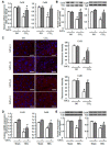
Cx32 and Cx26 expression in injured liver tissue with or without gadolinium chloride (GdCl3) administration. Four weeks after GdCl3 administration in CCl4-treated mice or two weeks after GdCl3 administration in bile duct ligation (BDL) mice, liver tissue was collected. Immuno-fluorescence staining, western blot and qRT-PCR analysis for the expression of Cx32 and Cx26 were performed. (A) Cx32 and Cx26 mRNA expression in CCl4-injured liver. (B) Cx32 and Cx26 protein expression in CCl4-injured liver. (C) Immunofluorescence staining for Cx32 and Cx26 in OO-treated, CCl4-treated, GdCl3 plus OO-treated and GdCl3 plus CCl4-treated liver. Scale bars, 20 μm. (D) Cx32 and Cx26 mRNA expression in BDL-injured liver. (E) Cx32 and Cx26 protein expression in BDL-injured liver. Typical autoradiograms are shown. Expression of tubulin was checked to correct for variations in protein loading and transfer. All results were confirmed in three independent experiments. Data are presented as the mean ± SEM, and were analyzed by ANOVA for analysis of variance. * p < 0.05 vs. OO-treated mice. # p < 0.05 vs. CCl4-treated mice without GdCl3 administration.
Figure 3
Figure 3
The gap junctional intercellular communication (GJIC) function in CCl4-treated mice with or without GdCl3 administration. (A) Digital images of Lucifer yellow dye transfer in liver treated with OO, CCl4, GdCl3 plus OO, GdCl3 plus CCl4. Scale bars, 100 μm. (B) GJIC function and integrity was determined by quantifying the distance of the Lucifer yellow dye transfer from the incision. Data are presented as the mean ± SEM and were analyzed by ANOVA for the analysis of variance. * p < 0.05 vs. OO-treated mice. # p < 0.05 vs. CCl4-treated mice without GdCl3 administration.
Figure 4
Figure 4
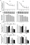
Cx32 and Cx26 expression in CCl4- or lipopolysaccharide (LPS)-injured primary hepatocytes with or without GdCl3 pretreatment. The expression of Cx32 and Cx26 mRNA (A) and protein (B) in primary hepatocytes which were treated with CCl4 of different concentrations. Cx32 and Cx26 mRNA expression in response to CCl4 (C) or LPS (D) with the pre-treatment of 200 μM GdCl3, 1 mM MgCl2 or 1 mM CaCl2 for 1 h, following 2 h of CCl4 or LPS treatment. All results were confirmed in three independent experiments. Data are presented as the mean ± SEM, and were analyzed by ANOVA for analysis of variance. * p < 0.05 vs. Ctrl. # p < 0.05 vs. CCl4-treated hepatocytes without GdCl3 administration.
Figure 5
Figure 5
The expression of Cx32 and Cx26 in CCl4-treated liver tissue with or without clodronate administration. Clodronate liposome (10 L/g B.W.) injection was performed once 72 h before CCl4 treatment to delete Kupffer cells. The qRT-PCR, western blot analysis and immunofluorescence staining for the expression of Cx32 and Cx26 were performed. (A) Cx32 and Cx26 mRNA expression in injured liver. (B) Cx32 and Cx26 protein expression in injured liver. Typical autoradiograms are shown. Expression of tubulin was checked to correct for variations in protein loading and transfer. (C) Immunofluorescence staining for Cx32 and Cx26 in injured liver. Scale bars, 100 μm. All results were confirmed in three independent experiments. Data are presented as the mean ± SEM, and were analyzed by ANOVA for analysis of variance. * p < 0.05 vs. Ctrl. # p < 0.05 vs. CCl4-treated mice alone.
Figure 6
Figure 6
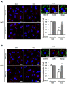
Immunofluorescence staining for Cx32 and Cx26 in CCl4-injured primary hepatocytes with or without GdCl3 pretreatment. Immunofluorescence staining for Cx32 (A) and Cx26 (B) in Ctrl-treated, CCl4-treated, GdCl3 plus CCl4-treated and GdCl3 plus CCl4-treated hepatocytes. Scale bars, 20 μm. The cell membrane was labeled with DiOC18, a membrane dye. Scale bars, 10 μm. DAPI was used to visualize nuclei (blue). Data are presented as the mean ± SEM, and were analyzed by ANOVA for the analysis of variance. * p < 0.05 vs. Ctrl. # p < 0.05 vs. CCl4-treated hepatocytes without GdCl3 administration.
Figure 7
Figure 7
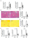
The effect of GdCl3 on liver inflammation and fibrosis in BDL mouse models. (A) Serum ALT and AST activity in BDL mice with or without GdCl3 administration. (B) Representative H&E-staining liver sections after GdCl3 administration in BDL liver. Scale bars, 50 µm. Inset: H&E-staining for Sham livers. (C) Quantification of inflammatory areas. (D) Representative images of Sirius Red staining after GdCl3 administration in BDL liver. Scale bars, 50 µm. Inset: Sirius Red staining for Sham livers. (E) Quantitative analysis of liver fibrosis. Ten randomly-selected fields were quantitated for each mouse using the Leica QWin V3 software. (F) Expression of Col α1(I), Col α1(III), and α-SMA mRNA levels in liver, measured by qRT-PCR. Data are presented as the mean ± SEM, and were analyzed by ANOVA for analysis of variance. ∗ p < 0.05 compared with the Sham group. # p < 0.05 compared with the BDL mice without GdCl3 administration.

Similar articles

Cited by

References

    1. Sánchez A., Castro C., Flores D.L., Gutiérrez E., Baldi P. Gap Junction Channels of Innexins and Connexins: Relations and Computational Perspectives. Int. J. Mol. Sci. 2019;20:2476. doi: 10.3390/ijms20102476. - DOI - PMC - PubMed
    1. Chanson M., Watanabe M., O’Shaughnessy E.M., Zoso A., Martin P.E. Connexin Communication Compartments and Wound Repair in Epithelial Tissue. Int. J. Mol. Sci. 2018;19:1354. doi: 10.3390/ijms19051354. - DOI - PMC - PubMed
    1. Oshima A. Structure and closure of connexin gap junction channels. FEBS Lett. 2014;588:1230–1237. doi: 10.1016/j.febslet.2014.01.042. - DOI - PubMed
    1. Maes M., Decrock E., Cogliati B., Oliveira A.G., Marques P.E., Dagli M.L.Z., Menezes G.B., Mennecier G., Leybaert L., Vanhaecke T., et al. Connexin and pannexin (hemi)channels in the liver. Front. Physiol. 2014;4:405. doi: 10.3389/fphys.2013.00405. - DOI - PMC - PubMed
    1. Yoshizawa T., Watanabe S., Hirose M., Miyazaki A., Sato N. Dimethylsulfoxide maintains intercellular communication by preserving the gap junctional protein connexin32 in primary cultured hepatocyte doublets from rats. J. Gastroenterol. Hepatol. 1997;12:325–330. doi: 10.1111/j.1440-1746.1997.tb00429.x. - DOI - PubMed

MeSH terms